. 2008 May 23;283(21):14295-308.
doi: 10.1074/jbc.M800501200. Epub 2008 Mar 10.

Functional characterization of murine interferon regulatory factor 5 (IRF-5) and its role in the innate antiviral response

Affiliations

Functional characterization of murine interferon regulatory factor 5 (IRF-5) and its role in the innate antiviral response

Andrea Paun et al. J Biol Chem. .

Abstract

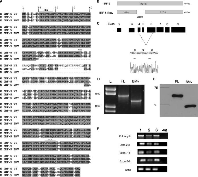
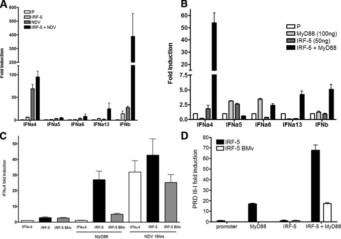
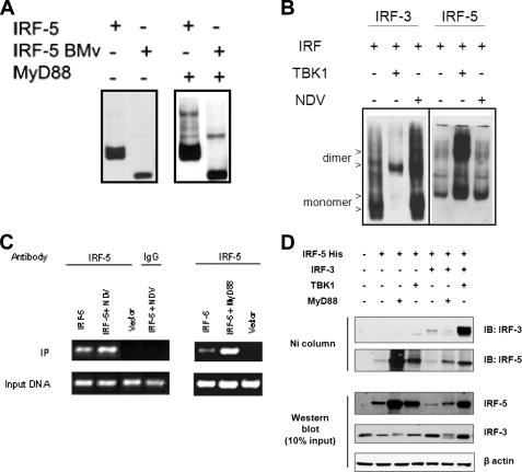
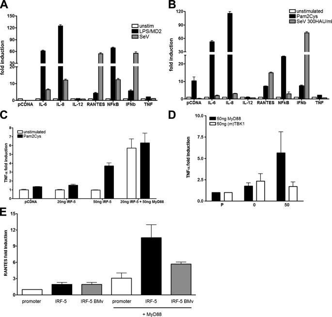
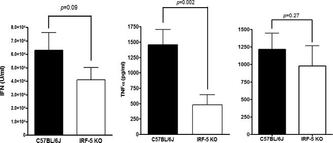
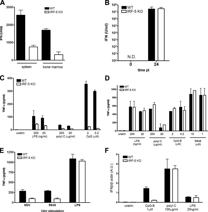
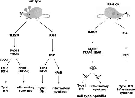
Although the role of human IRF-5 in antiviral and inflammatory responses in vitro has been well characterized, much remains to be elucidated about murine IRF-5. Murine IRF-5, unlike the heavily spliced human gene, is primarily expressed as a full-length transcript, with only a single splice variant that was detected in very low levels in the bone marrow of C57BL/6J mice. This bone marrow variant contains a 288-nucleotide deletion from exons 4-6 and exhibits impaired transcriptional activity. The murine IRF-5 can be activated by both TBK1 and MyD88 to form homodimers and bind to and activate transcription of type I interferon and inflammatory cytokine genes. The importance of IRF-5 in the antiviral and inflammatory response in vivo is highlighted by marked reductions in serum levels of type I interferon and tumor necrosis factor alpha (TNFalpha) in Newcastle disease virus-infected Irf5(-)(/)(-) mice. IRF-5 is critical for TLR3-, TLR4-, and TLR9-dependent induction of TNFalpha in CD11c(+) dendritic cells. In contrast, TLR9, but not TLR3/4-mediated induction of type I IFN transcription, is dependent on IRF-5 in these cells. In addition, IRF-5 regulates TNFalpha but not type I interferon gene transcription in Newcastle disease virus-infected peritoneal macrophages. Altogether, these data reveal the cell type-specific importance of IRF-5 in MyD88-mediated antiviral pathways and the widespread role of IRF-5 in the regulation of inflammatory cytokines.


Figures

FIGURE 1.
FIGURE 1.
Characterization of mouse IRF-5 and its splice variants. A, protein sequence alignment of human IRF-5 variant 5 with mouse IRF-5 and the mouse IRF-5 BMv. The deletion in IRF-5 BMv is shown schematically both for the protein (B) and the genomic DNA sequences (C). L, molecular weight markers (ladder). Expression of ectopic MuIRF-5 (full-length (FL)) and IRF-5 BMv proteins in transfected 293T cells was identified by immune blotting (D), and mRNA expression was identified by RT-PCR (E). F, to detect the possible presence of IRF-5 splice variants in C57BL/6J (lane 1), BALB/c (lane 2), and NZB mice (lane 3), regions representing full-length IRF-5 and distinct exons were amplified by two-step RT-PCR using total splenic RNA.
FIGURE 2.
FIGURE 2.
MuIRF-5 preferentially stimulates the murine IFNA4 gene promoter. A, 293T cells were transfected with the respective type I IFN promoter-luciferase reporter plasmids (50 ng), MuIRF-5 expressing plasmids (50 ng), or empty pcDNA6 vector and Renilla luciferase plasmid (10 ng). 20 h post-transfection cells were infected with NDV for 16 h and assayed for luciferase activity that was then normalized against Renilla luciferase. B, 293T cells were transfected with IRF-5 (50 ng) in the presence or absence of MyD88 (100 ng), with Renilla luciferase plasmid (10 ng) and type I IFN promoter-luciferase reporters (50 ng). C, 293T cells were transfected with IRF-5 full-length or IRF-5 BMv (100 ng) and MyD88 (50 ng) plasmids, IFNA4 reporter plasmid, and Renilla luciferase plasmid. Where indicated, cells were also infected with NDV for 16 h. D, 293T cells were transfected with IRF-5 full-length or IRF-5 BMv (10 ng), MyD88 (40 ng), PRDIII-I reporter plasmid, and Renilla luciferase plasmid. B and D, samples were assayed for luciferase activity 24 h post-transfection and normalized against Renilla luciferase. Data shown are combined from 2 or 3 independent experiments in A and C assays and triplicate repeats for B and D experiments.
FIGURE 3.
FIGURE 3.
MyD88-activated IRF-5 dimerizes and binds to the IFNA4 promoter. A, 293T cells were transfected with MyD88, full-length IRF-5, or IRF-5 BMv (all 0.5 μg) plasmids as indicated. Cells were lysed 24 h after transfection, proteins separated on a 7.5% native gel, and dimers detected by immunoblotting with anti-IRF-5 antibody. B, 293T cells were transfected as indicated with IRF-5 or IRF-3 and TBK1 plasmids (all 0.8 μg). 18 h after transfection cells were infected with NDV for 6 h. Proteins were analyzed on native and SDS-polyacrylamide gels and visualized by immunodetection. β-Actin levels were determined as a control for equal protein loading. C, in vivo binding of IRF-5 to the murine IFNA4 promoter was analyzed using the chromatin immunoprecipitation assay. The 293T cells were transfected with IFNA4 reporter (150 ng) and IRF-5 (110 ng) plasmids and either cotransfected with MyD88 plasmid (1.2 μg) or 18 h after transfection infected with NDV for 6 h. The chromatin immunoprecipitation assay was performed 24 h post-transfection as described under “Experimental Procedures.” Transfection with empty vector and immunoprecipitation with a nonspecific IgG antibody were used as controls. D, 293T cells (5 × 10⁷) were transfected with a combination of IRF-3, IRF-5, MyD88, and TBK-1 plasmids, and 24 h after the transfection, cells were lysed under nondenaturing conditions, and IRF-5 was purified on Ni+-charged resin as described under supplemental Experimental Procedures. IB, immunoblot.
FIGURE 4.
FIGURE 4.
Role of IRF-5 in TLR-dependent induction of inflammatory cytokines. 293T cells stably expressing TLR4 (A) or TLR2 (B) were transfected with luciferase reporters containing promoters of different inflammatory cytokines (40 ng), IRF-5 (100 ng), and Renilla luciferase (40 ng) plasmids. 8 h after transfection, cells were infected with Sendai virus (300 HAU/ml) or stimulated with Pam2Cys (10 nm) or LPS (10 ng/ml) in the presence of supernatants from MD2-expressing cells for 16 h. C, cells were transfected with the indicated amounts of IRF-5 and MyD88, Renilla luciferase (40 ng) plasmids, and TNFA reporter plasmid (40 ng) as described above. Transfected cells were left unstimulated or stimulated with 10 nm Pam2Cys for 16 h. D, 293T cells were transfected with IRF-5 (100 ng) and MyD88 or TBK1 (50 ng) plasmids, TNFA reporter, and Renilla luciferase plasmids. E, 293T cells were transfected as indicated with full-length IRF-5 or IRF-5 BMv (100 ng) and MyD88 (50 ng) plasmids, RANTES reporter plasmid, and Renilla luciferase plasmid. D and E samples were assayed for luciferase activity 24 h post-transfection and normalized against Renilla luciferase. Data shown are combined triplicates from two independent experiments.
FIGURE 5.
FIGURE 5.
Role of IRF-5 in the antiviral and inflammatory response to NDV infection in vivo. IRF-5-deficient mice (Irf5/) and C57BL6/J (wt) mice were infected with NDV (50 HAU) for 6 h and then the collected serum was analyzed for type I IFN by bioassay (left), TNFα (middle), and IL-6 by ELISA (right). Data shown are combined from four independent experiments, wild type n = 12 and Irf5–/– n = 15 mice.
FIGURE 6.
FIGURE 6.
Cell type-specific role of IRF-5 in the antiviral and inflammatory response. A, total white blood cells from spleen and bone marrow cells (2 × 10⁶ cells). B, peritoneal macrophages (2 × 10⁶ cells) from WT and Irf5/ mice were infected with NDV (50 HAU) for 24 h. The levels of type I IFN in the culture medium were measured by bioassay. C, peritoneal macrophages from WT and Irf5/ mice were infected with NDV (50 HAU) and treated with R848 (10 μm) or LPS (20 ng/ml) for 24 h, and TNFα levels in the supernatant were measured by ELISA. D, BMDC. E, purified splenic CD11c+ DC (1 × 10⁵ cells) from WT and Irf5/ mice were stimulated for 16 h with the respective TLR ligands, and levels of TNFα in the medium were measured by ELISA. F, purified splenic CD11c+ DC (1 × 10⁶ cells) from WT and Irf5/ mice were stimulated for 2 h with respective TLR ligands. Cells were harvested; total RNA was isolated, and relative levels of IFNβ were measured by real time PCR. Transcript levels are shown in arbitrary units (A.U.) compared with β-actin.
FIGURE 7.
FIGURE 7.
The role of IRF-5 in the innate antiviral response. Following infection, viral nuclei acids are recognized by membrane-bound TLR and cytoplasmic RIG-I/MDA5 receptors. Our data, in conjunction with data of others (32), have shown that IRF-5 is activated by TLR7- or TLR9-, MyD88-dependent pathway. Activated IRF-5 stimulates expression of IFNA and IFNB genes as well as the inflammatory cytokines. However, although the role of IRF-5 in the activation of type I IFN genes is restricted to the MyD88 pathway and to cells that express the TLR7 or TLR9 receptors, the activation of the inflammatory genes by TLR4 and TLR3 seems also to be dependent on IRF-5, and consequently the role of IRF-5 in the induction of these inflammatory cytokines is broader and less cell type-restricted. There is no evidence that IRF-5 plays a substantial, if any, role in the RigI, IPS1 (VISA/MAVS/CARDif) pathway, and whether it can be activated by the MAD5 pathways is yet to be determined.

References

    1. Akira, S., Uematsu, S., and Takeuchi, O. (2006) Cell 124 783–801 - PubMed
    1. O'Neill, L. A. (2006) Curr. Opin. Immunol. 18 3–9 - PubMed
    1. Hornung, V., Ellegast, J., Kim, S., Brzozka, K., Jung, A., Kato, H., Poeck, H., Akira, S., Conzelmann, K. K., Schlee, M., Endres, S., and Hartmann, G. (2006) Science 314 994–997 - PubMed
    1. Yoneyama, M., Kikuchi, M., Matsumoto, K., Imaizumi, T., Miyagishi, M., Taira, K., Foy, E., Loo, Y. M., Gale, M., Jr., Akira, S., Yonehara, S., Kato, A., and Fujita, T. (2005) J. Immunol. 175 2851–2858 - PubMed
    1. Yoneyama, M., Kikuchi, M., Natsukawa, T., Shinobu, N., Imaizumi, T., Miyagishi, M., Taira, K., Akira, S., and Fujita, T. (2004) Nat. Immunol. 5 730–737 - PubMed

Publication types

MeSH terms

Associated data