. 2008 May 16;283(20):13714-24.
doi: 10.1074/jbc.M709329200. Epub 2008 Mar 10.

Caveolin-1 up-regulation during epithelial to mesenchymal transition is mediated by focal adhesion kinase

Affiliations

Caveolin-1 up-regulation during epithelial to mesenchymal transition is mediated by focal adhesion kinase

Kelly M Bailey et al. J Biol Chem. .

Abstract

Emerging evidence has shown that caveolin-1 is up-regulated in a number of metastatic cancers and can influence various aspects of cell migration. However, in general, the role of caveolin-1 in cancer progression is poorly understood. In the present study, we examined alterations in caveolin-1 expression during epithelial-to-mesenchymal transition (EMT) and the ability of caveolin-1 to alter cancer cell adhesion, an aspect of cell motility. We employed two EMT cell models, the human embryonic carcinoma cell line NT2/D1, and TGF-beta1-treated NMuMG cells, which are derived from normal mouse mammary epithelia. Caveolin-1 expression was substantially up-regulated in both cell lines following the induction of EMT and was preceded by increased activation of focal adhesion kinase (FAK) and Src, two known tyrosine kinases involved in EMT. We hypothesized that caveolin-1 expression could be influenced by increased FAK phosphorylation, to which Src is a known contributor. Examination of FAK+/+ and FAK-/- mouse embryonic fibroblasts revealed that in cells devoid of FAK, caveolin-1 expression is strikingly diminished. Using FAK and superFAK constructs and the novel FAK inhibitor PF-228, we were able to demonstrate that indeed, FAK can regulate caveolin-1 expression. We also found that Src can contribute to increases in caveolin-1 expression, however, only in the presence of FAK. From the culmination of this data and our functional analyses, we conclude that caveolin-1 expression can be up-regulated during EMT, and further, once expressed, caveolin-1 can greatly influence cancer cell adhesion.


Figures
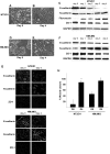
FIGURE 1.
FIGURE 1.
Stimulated NT2/D1 and NMuMG cell models both demonstrate epithelial to mesenchymal transition. A and B, morphology of unstimulated (day 0) and stimulated (day 4) NT2/D1 human embryonic carcinoma cells line NT2/D1 is shown in A and B, respectively. C, lysates from day 0- to day 4-stimulated NT2/D1 cells were subjected to Western blot analysis for (from top to bottom) E-cadherin, N-cadherin, fibronectin, and ZO-1. D and E, morphology of untreated (day 0) or 2 ng/ml TGF-β1-treated (day 4) NMuMG cells is shown in D and E, respectively. F, lysates from day 0- to day 4-stimulated NMuMG cells were subjected to Western blot analysis for E-cadherin, N-cadherin, and ZO-1. Immunoblots for GAPDH are included in C and F, and serve as equal protein loading controls. G, day 0 (unstimulated) and day 4 (stimulated) NT2/D1 (top panels) and NMuMG (bottom panels) were immunofluorescently labeled for E-cadherin, N-cadherin, and ZO-1. H, stimulated (D4) and unstimulated (D0) NT2/D1, and NMuMG cells were subjected to wound healing scratch assays and imaged over 10 h. The average % wound closure is graphed (*, D0 versus D4 NT2/D1, p value = 0.001; D0 versus D4, p value = 0.0001).
FIGURE 2.
FIGURE 2.
Caveolin-1 expression is up-regulated during EMT. A, Western blot analysis for caveolin-1 expression in day 0 to day4 NT2/D1 cell lysates. B, Western blot analysis for caveolin-1 in day 0 to day 4 NMuMG lysates. Immunoblots for GAPDH are included in A and B and serve as equal protein loading controls.
FIGURE 3.
FIGURE 3.
Phosphorylation of FAK and Src is increased during EMT. A, day 0 to day 4 NT2/D1 lysates were subjected to Western blot analysis for (from top to bottom) pFAK (Y397), pFAK (Y861), FAK, pSrc (Y418), Src, and GAPDH (an equal protein loading control). An identical Western panel in B shows the day 0 to day 4 NMuMG cell lysates.
FIGURE 4.
FIGURE 4.
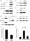
FAK is a critical mediator of caveolin-1 expression. A, FAK+/+ and FAK-/- MEF cell lysates were subjected to Western blot analysis for caveolin-1 (top) and FAK (middle). B, FAK-/- MEFs were transfected with FAK (GFP-tagged), superFAK, or mock-transfected. Lysates from these three groups were then subjected to Western blot analysis for caveolin-1 (top) and FAK (middle). Note the band for FAK in the FAK-GFP-transfected cells is shifted upward, reflecting the increase in molecular weight because of the presence of GFP. C, FAK+/+ MEFs were treated with PF-228, an inhibitor of FAK catalytic activity, using the concentrations indicated for 24 h. Lysates were made and subjected to Western blot analysis for caveolin-1, pFAK(Y397), and FAK. D, NT2/D1 cells stimulated for 2 days were treated with PF-228 using the concentrations indicated. NT2/D1 lysates were made at day 3 and then subjected to Western blot analysis for caveolin-1, pFAK (Y397), and FAK. Immunoblots for GAPDH are included in A–D and serve as equal protein loading controls. Relative expression values of caveolin-1 are included below the blots in C and D and represent caveolin-1/normalized GAPDH signal obtained by densitometry analysis.
FIGURE 5.
FIGURE 5.
Src-induced caveolin-1 expression is FAK-dependent. A, SYF-/- MEFs were nucleofected with the active Src construct c-SrcY527F or mock-nucleofected. Lysates were subjected to Western blot analysis for caveolin-1, pSrc (Y418), pFAK (Y397), pFAK (Y861), and FAK. B, FAK-/- MEFs were nucleofected with c-SrcY527F or mock-nucleofected. These lysates and a FAK+/+ lysate control were subjected to Western blot analysis for caveolin-1, pSrc (Y418), and FAK. C, SYF-/- MEFs were nucleofected with superFAK or mock-nucleofected, and lysates were analyzed via Western blot analysis for alterations in caveolin-1 expression. D, NT2/D1 stimulated for 2 days were treated with 5 μm PP2 for 24 h, and the cells (day 3) were then lysed and analyzed for caveolin-1 expression via Western blot analysis. E, NT2/D1 cells were stimulated as in D followed by treatment with SU-6656 or controls, and lysates were subjected to Western blot analysis for caveolin-1. F, total RNA was isolated from FAK+/+ and FAK-/- MEFs. The relative amount of caveolin-1 mRNA was measured by quantitative real-time PCR analysis normalized against mouse 18 S (see “Experimental Procedures”). *, p = 0.004. G, FAK-/- MEFs were nucleofected with superFAK, c-SrcY527F, or mock-nucleofected. RNA was isolated from each of these three groups, and caveolin-1 mRNA was analyzed as in F. **, p = 0.001. Real-time experiments in F and G were performed at least in triplicate. GAPDH control blots for equal protein loading are included in A–E.
FIGURE 6.
FIGURE 6.
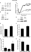
Down-regulation of caveolin-1 affects cell attachment, adhesion, and migration in H1703 human lung cancer cells. A, H1703 cells were treated with increasing concentrations of PF-228 or control (DMSO only) and blotted for pFAK (Y397) and caveolin-1. B, H1703 cells were treated with 5 μm PP2 or control (DMSO only) and blotted for pFAK (Y397) and caveolin-1. C, control or caveolin-1 siRNA-treated H1703 cells were examined for alterations in attachment resistance using an ECIS attachment assay. Targeted knockdown of caveolin-1 using caveolin-1 siRNA (black curve) lead to a significant increase in resistance compared with that of the control (gray curve) (p < 0.01). D, some of the control or siRNA-treated cells from C were lysed and immunoblotted with antibody for caveolin-1. E, control or caveolin-1 siRNA-treated H1703 cells were immunofluorescently labeled for actin and paxillin, randomly imaged, and relative cell spreading areas were quantified using Image J software. Targeted knockdown of caveolin-1 increases the total cell spreading area compared with cells treated with control siRNA. *, p < 0.05. F, total number of focal adhesions in either control or caveolin-1-treated cells was quantified using the same images in E. Note that the knockdown of caveolin-1 leads to an increase in the total number of focal adhesions in the cell. *, p < 0.05. G, to determine whether adhesion numbers were affected by cell spreading area, cells were arbitrarily subdivided into small, medium, and large groups. Bars represent the relative fold increase in focal adhesions in caveolin-1 siRNA-treated cells over that of control siRNA-treated cells (normalized to a value of 1) for each group. *, p < 0.05. H, control or caveolin-1 siRNA-treated cells were subjected to Boyden chamber migration assays. Knockdown of caveolin-1 significantly decreases cell migration compared with that of controls (*, p = 0.001). GAPDH blots were included as equal protein loading controls in A, B, and D.

References

    1. Rothberg, K. G., Heuser, J. E., Donzell, W. C., Ying, Y. S., Glenney, J. R., and Anderson, R. G. (1992) Cell 68 673-682 - PubMed
    1. Glenney, J. R., Jr. (1989) J. Biol. Chem. 264 20163-20166 - PubMed
    1. Glenney, J. R., Jr., and Zokas, L. (1989) J. Cell Biol. 108 2401-2408 - PMC - PubMed
    1. Williams, T. M., and Lisanti, M. P. (2005) Am. J. Physiol. Cell Physiol. 288 C494-C506 - PubMed
    1. Hayashi, K., Matsuda, S., Machida, K., Yamamoto, T., Fukuda, Y., Nimura, Y., Hayakawa, T., and Hamaguchi, M. (2001) Cancer Res. 61 2361-2364 - PubMed

Publication types

Substances