. 2008 Mar 17;205(3):725-35.
doi: 10.1084/jem.20072525. Epub 2008 Mar 10.

CD94-NKG2A recognition of human leukocyte antigen (HLA)-E bound to an HLA class I leader sequence

Affiliations

CD94-NKG2A recognition of human leukocyte antigen (HLA)-E bound to an HLA class I leader sequence

Emma J Petrie et al. J Exp Med. .

Abstract

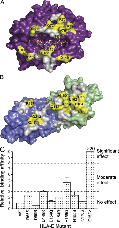
The recognition of human leukocyte antigen (HLA)-E by the heterodimeric CD94-NKG2 natural killer (NK) receptor family is a central innate mechanism by which NK cells monitor the expression of other HLA molecules, yet the structural basis of this highly specific interaction is unclear. Here, we describe the crystal structure of CD94-NKG2A in complex with HLA-E bound to a peptide derived from the leader sequence of HLA-G. The CD94 subunit dominated the interaction with HLA-E, whereas the NKG2A subunit was more peripheral to the interface. Moreover, the invariant CD94 subunit dominated the peptide-mediated contacts, albeit with poor surface and chemical complementarity. This unusual binding mode was consistent with mutagenesis data at the CD94-NKG2A-HLA-E interface. There were few conformational changes in either CD94-NKG2A or HLA-E upon ligation, and such a "lock and key" interaction is typical of innate receptor-ligand interactions. Nevertheless, the structure also provided insight into how this interaction can be modulated by subtle changes in the peptide ligand or by the pairing of CD94 with other members of the NKG2 family. Differences in the docking strategies used by the NKG2D and CD94-NKG2A receptors provided a basis for understanding the promiscuous nature of ligand recognition by NKG2D compared with the fidelity of the CD94-NKG2 receptors.


Figures

Figure 1.
Figure 1.
The CD94-NKG2A–HLA-EVMAPRTLFL complex. NKG2A and CD94 are represented as blue and pale green ribbon structures, respectively. The heavy chain of HLA-E and β2m are shown as violet and cyan ribbons, respectively, with the VMAPRTLFL peptide in orange sticks. (A) Side view of CD94-NKG2A docking onto HLA-EVMAPRTLFL. (B) Top view of CD94-NKG2A docking onto the surface of HLA-EVMAPRTLFL.
Figure 2.
Figure 2.
Interaction of CD94-NKG2A subunits to HLA-EVMAPRTLFL. (A) Electrostatics of CD94-NKG2A and HLA-E (represented as surface displaying charged regions) reveal charge complementarity that drives the interaction. (B) CD94-NKG2A interaction footprint on HLA-EVMAPRTLFL. The surface of HLA-E is shown with the peptide as orange space fill. Contacts to HLA-E made by CD94 are represented in pale green, and those made by NKG2A are in blue. Both subunits contact P5-Arg and are shown in teal. (C) Key interactions of CD94 to HLA-E α1 helix. Hydrogen bonds are formed between Gln113 to Asp69 of HLA-E, which also forms a salt bridge with Arg171 of CD94. Asn170 and Glu164 both form hydrogen bonds with Gln72 of HLA-E, whereas Glu89 forms a hydrogen bond with Thr146. (D) Binding of CD94 to HLA-E involves hydrophobic interactions between CD94 Phe114 and Leu162 by Ile73 and Val76 of HLA-E and P8-Phe of the peptide. Leu162 is also involved in VDW interactions further along the α1 helix. In addition, neighboring Asp163 forms a salt bridge with Arg75 of HLA-E, and Ser143 of CD94 forms a hydrogen bond with Arg79. (E) Key NKG2A interactions with HLA-E α2 helix. Arg137 forms a hydrogen bond with Ser151 and a salt bridge with Glu154 of HLA-E. His155 of HLA-E forms a hydrogen bond with Ser172 while making VDW contacts with Pro171. Ala158 of HLA-E makes VDW contacts with Gln212 and Lys217 of NKG2A, the latter of which forms a salt bridge with Asp162 and the α2 helix. Hydrogen bonds are represented as black dashed lines, and salt bridges are in blue.
Figure 3.
Figure 3.
Peptide-mediated contacts to CD94-NKG2A. (A) The surface of CD94 (pale green) clearly accommodates Arg5 and Phe8 of the peptide (orange sticks) presented on HLA-E (violet ribbon). NKG2A (blue surface) makes minimal contact with the peptide. (B) Pro171 is the only residue of NKG2A that interacts with the peptide, at P5-Arg. CD94 dominates recognition of the peptide making several interactions, including hydrogen bonds of Ser110 to P5-Arg and Gln112 to P6-Thr. A distinct polar pocket created by Asn156, Asn158, Asn160, and Phe114 on CD94 accommodates P8-Phe. (C) KK50.4 TCR interactions to HLA-E–presenting peptide VMAPRTLIL. Interactions are dominated by the Vβ chain of the TCR in a manner analogous to CD94-NKG2A interactions.
Figure 4.
Figure 4.
Energetically important residues and the corresponding structural footprint. (A) Surface representation of HLA-E with bound peptide (orange). (B) Surface representation of CD94-NKG2A, with CD94 colored green and NKG2A colored blue. Residues that have been shown, via mutagenesis, to influence binding (moderate and significant effects) are shown in yellow. Residues that make contacts but have no demonstrable effect on the affinity of binding are shown in gray. (C) Relative binding of HLA-E mutants to CD94-NKG2A. The binding affinity of each mutant was determined by surface plasmon resonance, and the relative effect on binding was compared with wild-type (WT) HLA-E, which was normalized to a value of 1. There was no effect on binding if there was a less than threefold difference in binding affinity, whereas a greater than eightfold difference represented a significant effect.
Figure 5.
Figure 5.
Comparison of polar versus nonpolar interactions by CD94-NKG2A and NKG2D homodimer. Nonpolar interactions are represented in purple, and polar interactions are in blue. In situations where a residue makes both nonpolar interactions as well either a salt bridge or hydrogen bond, it was represented as making a polar interaction. (A) Ribbon representation of CD94-NKG2A in complex with HLA-EVMAPRTLFL and NKG2D homodimers binding to MICA and ULBP3. The subunit of the NKG2D homodimer equivalent to CD94 is represented in pale green, and the subunit equivalent to NKG2A is in blue. The MHC-like molecules, MICA and ULBP3, do not associated with β2m (represented in cyan), and these molecules are represented in violet. (B) “Polarity” footprints onto MHC molecules HLA-E by CD94-NKG2A, and MICA and ULPB3 by NKG2D homodimer. The peptide presented by HLA-E is represented in orange space fill. (C) Residues of CD94-NKG2A and the NKG2D homodimer involved in polar and nonpolar interactions.

References

    1. Moretta, L., C. Bottino, D. Pende, R. Castriconi, M.C. Mingari, and A. Moretta. 2006. Surface NK receptors and their ligands on tumor cells. Semin. Immunol. 18:151–158. - PubMed
    1. Boyton, R.J., and D.M. Altmann. 2007. Natural killer cells, killer immunoglobulin-like receptors and human leucocyte antigen class I in disease. Clin. Exp. Immunol. 149:1–8. - PMC - PubMed
    1. Cella, M., H. Nakajima, F. Facchetti, T. Hoffmann, and M. Colonna. 2000. ILT receptors at the interface between lymphoid and myeloid cells. Curr. Top. Microbiol. Immunol. 251:161–166. - PubMed
    1. Natarajan, K., N. Dimasi, J. Wang, D.H. Margulies, and R.A. Mariuzza. 2002. MHC class I recognition by Ly49 natural killer cell receptors. Mol. Immunol. 38:1023–1027. - PubMed
    1. Gasser, S., and D.H. Raulet. 2006. Activation and self-tolerance of natural killer cells. Immunol. Rev. 214:130–142. - PubMed

Publication types

MeSH terms