. 2008 May;76(5):1848-57.
doi: 10.1128/IAI.01424-07. Epub 2008 Mar 10.

A novel Treponema pallidum antigen, TP0136, is an outer membrane protein that binds human fibronectin

Affiliations

A novel Treponema pallidum antigen, TP0136, is an outer membrane protein that binds human fibronectin

Mary Beth Brinkman et al. Infect Immun. 2008 May.

Abstract

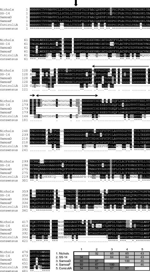
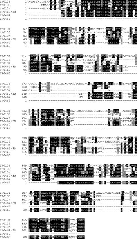
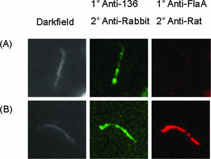
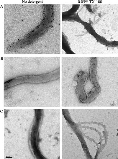
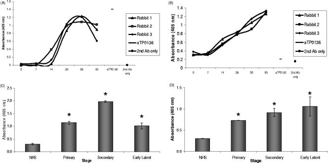
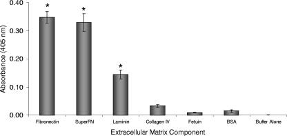
The antigenicity, structural location, and function of the predicted lipoprotein TP0136 of Treponema pallidum subsp. pallidum were investigated based on previous screening studies indicating that anti-TP0136 antibodies are present in the sera of syphilis patients and experimentally infected rabbits. Recombinant TP0136 (rTP0136) protein was purified and shown to be strongly antigenic during human and experimental rabbit infection. The TP0136 protein was exposed on the surface of the bacterial outer membrane and bound to the host extracellular matrix glycoproteins fibronectin and laminin. In addition, the TP0136 open reading frame was shown to be highly polymorphic among T. pallidum subspecies and strains at the nucleotide and amino acid levels. Finally, the ability of rTP0136 protein to act as a protective antigen to subsequent challenge with infectious T. pallidum in the rabbit model of infection was assessed. Immunization with rTP0136 delayed ulceration but did not prevent infection or the formation of lesions. These results demonstrate that TP0136 is expressed on the outer membrane of the treponeme during infection and may be involved in attachment to host extracellular matrix components.


Figures

FIG. 1.
FIG. 1.
Amino acid sequence heterogeneity of TP0136 among strains of Treponema. Sequences are identified by strain name, and the strains included are two syphilis-causing strains (T. pallidum subsp. pallidum Nichols and T. pallidum subsp. pallidum Street strain 14), two yaws strains (T. pallidum subsp. pertenue Samoa D and T. pallidum subsp. pertenue Samoa F), and the rabbit pathogen T. paraluiscuniculi Cuniculi A. The vertical arrow indicates the predicted signal sequence cleavage site, and a repeat region is indicated by horizontal arrows. Black shading indicates 50% sequence identity, gray shading indicates >50% functionally similar amino acids, and no shading indicates <50% sequence similarity. Percent identity (light gray) and percent similarity (dark gray) for each sequence are indicated in the inset table at bottom right. -, gap introduced to optimize alignment. The consensus line contains the following annotations: *, identical amino acid residues; functionally similar amino acid residues. Numbers on the left indicate the positions of the amino acid residues.
FIG. 2.
FIG. 2.
Sequence alignment of TP0136 paralogs indicated by genome sequence. Resequencing revealed a discrepancy with the published genome sequence that when translated yields the sequence TP0462/3R. Black shading indicates 100% sequence identity, gray shading indicates 75% sequence identity, and no shading indicates <50% sequence identity. -, gap introduced to optimize alignment. Numbers on the left indicate the positions of the amino acid residues.
FIG. 3.
FIG. 3.
Double label immunofluorescence microscopy following incubation with anti-rTP0136 and anti-FlaA antibodies with Percoll-purified T. pallidum in the absence (A) or presence (B) of 0.1% Triton X-100 detergent. Antibody binding of anti-rTP0136 antibody was detected using Alexa Fluor 488 goat anti-rabbit IgG (green), and antibody binding of anti-FlaA was detected using Alexa Fluor 546 goat anti-rat IgG (red).
FIG. 4.
FIG. 4.
Immunoelectron microscopy on T. pallidum cells following incubation with or without detergent and immunostaining with anti-rTP0136 antibody (A), the preimmune serum negative control (B), or anti-FlaA (C), followed by incubation with secondary antibody conjugated with gold particles. Bar length is 0.1 μm. TX-100, Triton X-100.
FIG. 5.
FIG. 5.
Reactivity of serum antibodies (Ab) from experimentally infected rabbits and human syphilis patients with purified rTP0136 protein, as determined by ELISA. (A) Reactivity of sera from individual rabbits (each indicated by a separate line) collected at sequential time points during an experimental T. pallidum infection with rTP0136 protein. Prechallenge rabbit sera (zero time) were used as a negative control, and anti-rTP0136 (aTP0136) antibody was used as a positive control for ELISA well coating. (B) Reactivity of individual rabbit sera to a protein lysate of T. pallidum was used as a control for the sensitivity of the rabbit sera used for panel A. As in panel A, prechallenge rabbit sera (zero time) were used as a negative control and anti-rTP0136 (aTP0136) was used as a positive control for plate coating. (C) Purified rTP0136 reactivity with immunoglobulin from sera pooled from patients with primary, secondary, and early latent syphilis. NHS were used as a negative control. (D) Reactivity of pooled human sera to the T. pallidum protein lysate was used as a positive control for the reactivity of the sera used for panel C. As in panel C, NHS were used as a negative control. Human serum samples were diluted 1:200, and rabbit samples were diluted 1:100. Statistical significance (P < 0.005) in comparison with negative controls (NHS) or prechallenge sera by the Student two-tailed t test is indicated by an asterisk.
FIG. 6.
FIG. 6.
Binding of purified rTP0136 protein with purified ECM proteins as measured by ELISA. The non-ECM proteins fetuin and BSA were used as negative controls. Statistical significance (P < 0.005) in comparison with binding to the negative control BSA by the Student two-tailed t test is indicated by an asterisk.

References

    1. Baker-Zander, S. A., E. W. Hook III, P. Bonin, H. H. Handsfield, and S. A. Lukehart. 1985. Antigens of Treponema pallidum recognized by IgG and IgM antibodies during syphilis in humans. J. Infect. Dis. 151264-272. - PubMed
    1. Baseman, J. B., J. C. Nichols, J. W. Rumpp, and N. S. Hayes. 1974. Purification of Treponema pallidum from infected rabbit tissue: resolution into two treponemal populations. Infect. Immun. 101062-1067. - PMC - PubMed
    1. Blanco, D. R., C. I. Champion, A. Dooley, D. L. Cox, J. P. Whitelegge, K. Faull, and M. A. Lovett. 2005. A monoclonal antibody that conveys in vitro killing and partial protection in experimental syphilis binds a phosphorylcholine surface epitope of Treponema pallidum. Infect. Immun. 733083-3095. - PMC - PubMed
    1. Brinkman, M. B., M. McKevitt, M. McLoughlin, C. Perez, J. Howell, G. M. Weinstock, S. J. Norris, and T. Palzkill. 2006. Reactivity of antibodies from syphilis patients to a protein array representing the Treponema pallidum proteome. J. Clin. Microbiol. 44888-891. - PMC - PubMed
    1. Cameron, C. 2006. The T. pallidum outer membrane and outer membrane proteins, p. 237-266. In J. D. Radolf and S. Lukehart (ed.), Pathogenic Treponema. Caister Academic Press, Norfolk, VA.

Publication types

MeSH terms

LinkOut - more resources