. 2008 Mar 10;180(5):1005-19.
doi: 10.1083/jcb.200707191.

Myoblasts and macrophages share molecular components that contribute to cell-cell fusion

Affiliations

Myoblasts and macrophages share molecular components that contribute to cell-cell fusion

Kostandin V Pajcini et al. J Cell Biol. .

Abstract

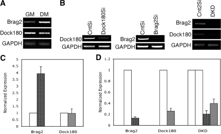
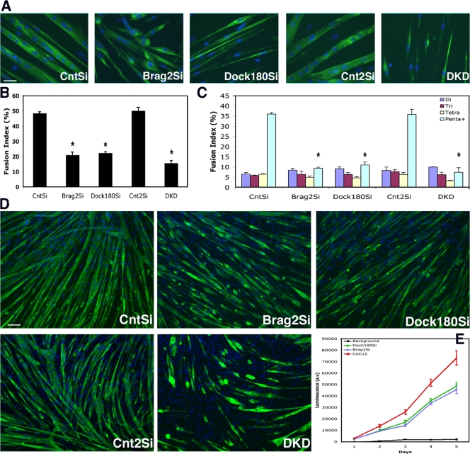
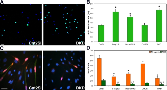
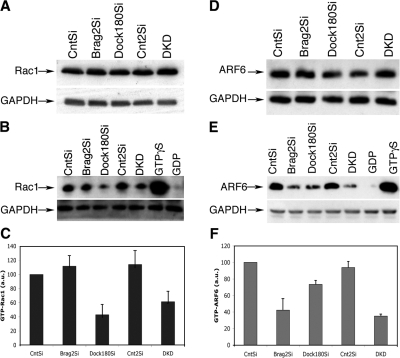
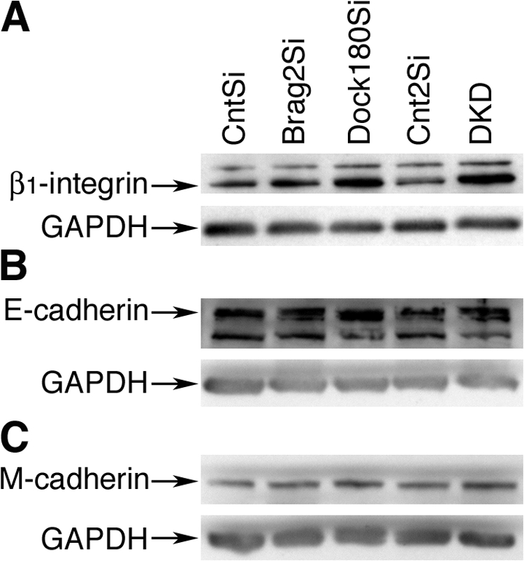
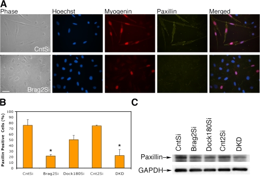
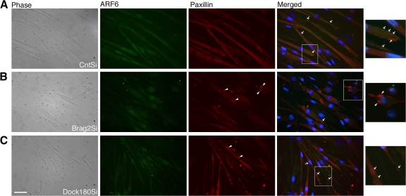
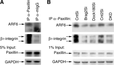
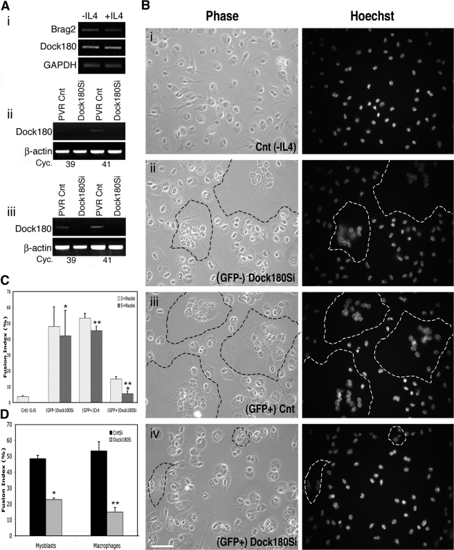
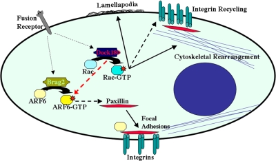
Cell-cell fusion is critical to the normal development of certain tissues, yet the nature and degree of conservation of the underlying molecular components remains largely unknown. Here we show that the two guanine-nucleotide exchange factors Brag2 and Dock180 have evolutionarily conserved functions in the fusion of mammalian myoblasts. Their effects on muscle cell formation are distinct and are a result of the activation of the GTPases ARF6 and Rac, respectively. Inhibition of ARF6 activity results in a lack of physical association between paxillin and beta(1)-integrin, and disruption of paxillin transport to sites of focal adhesion. We show that fusion machinery is conserved among distinct cell types because Dock180 deficiency prevented fusion of macrophages and the formation of multinucleated giant cells. Our results are the first to demonstrate a role for a single protein in the fusion of two different cell types, and provide novel mechanistic insight into the function of GEFs in the morphological maturation of multinucleated cells.


Figures

Figure 1.
Figure 1.
Expression and knockdown of Brag2 and Dock180 in C2C12 myoblasts. (A) Brag2 (1,100 bp) and Dock180 (2,485 bp) expression during growth (GM) and differentiation (DM) as shown by RT-PCR. GAPDH (250 bp). (B) Total RNA from cell lines Brag2Si, Dock180Si, and DKD, was harvested after d 4 in DM, and tested for Brag2 and Dock180 expression by RT-PCR. (C) Quantitative PCR analysis of Brag2 (dark gray) and Dock180 (light gray) expression in GM and d 4 DM. Values are normalized to the expression in CntSi cells (white). (D) Quantitative PCR analysis of Brag2 and Dock180 expression from Brag2Si, Dock180Si, and DKD cells. Values are normalized to the expression of CntSi and Cnt2Si cells. Error bars indicate the mean ± SE of three independent determinations.
Figure 2.
Figure 2.
Brag2- and Dock180-deficient cells have similar fusion index but disparate myotube morphologies. (A) Immunofluorescence images from each cell line, after d 6 in DM. Cells were labeled with primary antibody to MHC and secondary Alexa 488 (green) and Hoechst 33258 (blue). Bar, 50 μm. (B) Total fusion index analysis representing the number of nuclei in multinucleated myotubes divided by total number of nuclei in a field, with a myotube defined by at least three nuclei. A minimum of 4,000 nuclei were counted from random fields of each line at d 6 in DM. P value was determined with a t test, in which CntSi served as control for Brag2Si and Dock180Si cells and Cnt2Si served as control for DKD (*, P < 0.00001). (C) Fusion index analysis indicating the formation of di-, tri-, tetra-, or penta+ multinucleated myotubes after d 6 in DM. All error bars indicate the mean ± SE of at least four independent determinations (*, P < 0.00001). (D) Representative immunofluorescence images of d 6 DM fields used in the fusion index analysis. Bar, 200 μm. (E) β-gal complementation assay of fusion of Brag2Si, Dock180Si, and C2C12 control cells containing equal populations of α- and ω-fragments over the course of 5 d in DM. Background luminescence determined by C2C12 control cells containing α- and ω-fragments of β-gal seeded under proliferation conditions. (n = 3).
Figure 3.
Figure 3.
Delayed cell cycle withdrawal and differentiation in Brag2- and Dock180-deficient myoblasts. (A) Merged laser-scanning confocal image of representative fields of Cnt2Si and DKD myoblasts after 24 h in DM, labeled for BrdU (green) and with To-Pro (blue). BrdU+ nuclei are colored in teal in the merged image. Bar, 100 μm. (B) BrdU incorporation for each stable cell line after 24 h in DM and a 6-h BrdU incubation. (*P < 0.001). (C) Immunofluorescence of representative image fields of Cnt2Si and DKD myoblasts after 72 h in DM, labeled for myogenin (red), MHC (green), and with Hoechst 33258 (blue). Bar, 50 μm. (D) Differentiation as measured by myogenin (*, P < 0.0006) and MHC (**, P < 0.003; ***, P < 0.03) expression for each stable cell line after 72 h in DM. In all cases, error bars indicate the mean ± SE of three independent determinations, in which a minimum of 1,000 nuclei were counted per trial.
Figure 4.
Figure 4.
Brag2 and Dock180 deficiency decreases GTPase activity. (A) Western blot analysis of total expression of Rac in each cell line after d 3 in DM. Rac migrates as a 21-kD protein and GAPDH migrates as 35-kD protein. (B) Western blot indicating the level of activated Rac, as detected in lysates at d 3 in DM using a pull-down assay with GST-fused PAK protein-binding domain. Lysates from CntSi cell line were treated with GTPγS and GDP for positive and negative controls, respectively. (C) The histogram represents the levels of GTP-Rac as determined by three independent experiments. All samples were normalized to total protein and Brag2Si, Dock180Si, Cnt2Si, and DKD values are also normalized to the Rac-GTP levels observed in CntSi. (D) Western blot analysis of total expression of ARF6 after d 3 in DM. ARF6 migrates as a 20-kD protein. (E) Western blot indicating the level of activated ARF6, as detected in lysates at d 3 in DM using a pull-down assay with GST-fused GGA3 protein-binding domain, which specifically binds ARF6-GTP. (F) The histogram represents the levels of ARF6-GTP as determined by three independent experiments. All samples were normalized to total protein and Brag2Si, Dock180Si, Cnt2Si, and DKD values are also normalized to the ARF6-GTP levels observed in CntSi at d 3 in DM.
Figure 5.
Figure 5.
Dock180 deficiency in myotubes increases cellular levels of β1-integrin. (A) Western blot analysis of total protein levels of β1-integrin at d 6 in DM. β1-integrin migrates as a 130-kD protein and loading control GAPDH migrates as 35-kD protein. (B) Western blot analysis of total protein levels of E-cadherin. E-cadherin migrates as a 120-kD protein; the 90-kD band likely reflects digestion of the extracellular domain during lysate preparation. (C) Western blot analysis of M-cadherin total protein levels. M-cadherin migrates as a 130-kD protein.
Figure 6.
Figure 6.
Brag2-deficient myoblasts exhibit aberrant paxillin localization during differentiation. (A) Representative images of CntSi and Brag2Si myoblasts after d 3 in DM depicted in phase contrast and immunofluorescence, labeled with Hoechst 33258 (blue), myogenin (red), paxillin (yellow), and merged. Bar, 50 μm. (B) Paxillin redistribution in myogenin-positive cells, d 3 in DM. At least 1,000 myogenin-positive nuclei were counted for each experimental trial, and the error bars indicate the mean ± SE of three independent determinations (*, P < 0.01). (C) Western blot analysis of paxillin expression after d 5 in DM under fusion conditions. Paxillin migrates as a 65-kD protein.
Figure 7.
Figure 7.
ARF6 and paxillin fail to colocalize during early fusion in Brag2-deficient cells. (A) Representative images of CntSi, (B) Brag2Si, and (C) Dock180Si myoblasts after d 3 in DM depicted in phase contrast and immunofluorescence, labeled for ARF6 (green), paxillin (red), and merged with Hoechst 33258 (blue). Arrowheads point to clusters of paxillin observed in Brag2 and to a lesser extent in Dock180 knockdowns. Arrows point to colocalization sites of ARF6 and paxillin in the merged fields. Bar, 50 μm.
Figure 8.
Figure 8.
Paxillin physical interaction with ARF6 and β1-integrin is disrupted in Brag2 and Dock180-deficient cells. (A) Cell lysates of CntSi myoblasts in d 3 DM under fusion conditions were immunoprecipitated using anti-Paxillin antibody and mouse IgG serum as negative control. Immunoblotting was performed for ARF6 (20-kD) and β1-integrin (130- and 100-kD). 5% of lysate extract was immunoblotted for paxillin (65-kD) and GAPDH (35-kD) in order to verify equal amounts of cell lysates. (B) Cell lysates of each cell line were immunoprecipitated using anti-Paxillin antibody and immunoblotted for presence of ARF6 and β1-integrin. 1% of lysate extract was immunoblotted for paxillin and GAPDH in order to verify equal amounts of cell lysates.
Figure 9.
Figure 9.
Macrophages deficient in Dock180 fail to form MNGCs. (A) (i) Brag2 (1,100 bp) and Dock180 (745 bp) expression in thioglycollate-induced IP-harvested macrophages before and after treatment with IL-4 as indicated by RT-PCR analysis. β-actin (300 bp). (ii) Semi-quantitative RT-PCR showing expression of Dock180 in CD11b+Gr1+ monocytes obtained from GFP+ peripheral blood of PVR-Cnt-BM and PVR-Dock180Si-BM mice 4 wk after transplantation. (iii) Semi-quantitative RT-PCR showing expression of Dock180 in CD11b+ IP-macrophages of PVR-Cnt-BM and PVR-Dock180Si-BM mice 6 wk after transplantation under fusion conditions. (B) Phase-contrast and immunofluorescence images of IP-harvested macrophages from: (i) PVR-Cnt-BM macrophages serving as negative fusion control cultured without IL-4; (ii) GFP cells from PVR-Dock180Si-BM representing the endogenous macrophage population cultured with IL-4; (iii) GFP+ cells from PVR-Cnt-BM representing the transplanted control population cultured with IL-4; and (iv) GFP+ cells from PVR-Dock180Si-BM representing the transplanted Dock180-deficient population cultured with IL-4. MNGC formation can be distinguished by the cluster of nuclei with a large surrounding area of cytoplasm, the extent of which is indicated by dashed lines. (C) Fusion index analysis in macrophages from each of the four populations shown in B. Two multinucleated categories are analyzed and a minimum of 1,000 nuclei were counted for each fusion assay. Error bars indicate the mean ± SE of four independent fusion assays. P values were determined by t test between the two positive fusion controls and the PVR-Dock180Si-BM macrophages (*, P < 0.03; **, P < 0.0005). (D) Comparison of fusion indexes, of three or more nuclei, in myoblasts and macrophages deficient in Dock180. Error bars indicate the mean ± SE of four independent fusion assays (*, P < 0.00001; **, P < 0.0005).
Figure 10.
Figure 10.
Model for the roles of Brag2 and Dock180 in cell–cell fusion. Upon receipt of signals from fusion receptors on the cell surface Brag2 activates ARF6, which in turn transports paxillin to sites of focal adhesion, where it complexes with integrins thereby maintaining the structural integrity required during myotube maturation. Dock180 regulates Rac-GTP activity thus primarily contributing to lamellipodia formation and cytoskeletal rearrangement. Dock180 can activate ARF6, thus in part functioning in paxillin transport and possibly integrin recycling.

References

    1. Albert, M.L., J.I. Kim, and R.B. Birge. 2000. alphavbeta5 integrin recruits the CrkII-Dock180-rac1 complex for phagocytosis of apoptotic cells. Nat. Cell Biol. 2:899–905. - PubMed
    1. Boshans, R.L., S. Szanto, L. van Aelst, and C. D'Souza-Schorey. 2000. ADP-ribosylation factor 6 regulates actin cytoskeleton remodeling in coordination with Rac1 and RhoA. Mol. Cell. Biol. 20:3685–3694. - PMC - PubMed
    1. Brown, M.C., and C.E. Turner. 2004. Paxillin: adapting to change. Physiol. Rev. 84:1315–1339. - PubMed
    1. Charlton, C.A., W.A. Mohler, and H.M. Blau. 2000. Neural cell adhesion molecule (NCAM) and myoblast fusion. Dev. Biol. 221:112–119. - PubMed
    1. Charrasse, S., F. Comunale, M. Fortier, E. Portales-Casamar, A. Debant, and C. Gauthier-Rouviere. 2007. M-Cadherin activates Rac1 GTPase through the Rho-GEF trio during myoblast fusion. Mol. Biol. Cell. 18:1734–1743. - PMC - PubMed

Publication types

MeSH terms