. 2008 Mar 18;105(11):4168-72.
doi: 10.1073/pnas.0710285105. Epub 2008 Mar 10.

Specific requirement of the chromatin modifier mSin3B in cell cycle exit and cellular differentiation

Affiliations

Specific requirement of the chromatin modifier mSin3B in cell cycle exit and cellular differentiation

Gregory David et al. Proc Natl Acad Sci U S A. .

Abstract

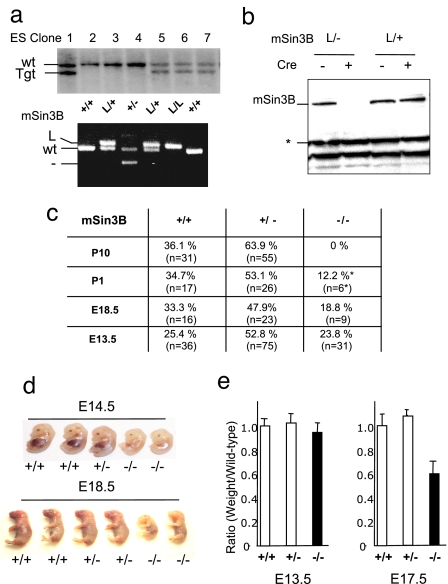
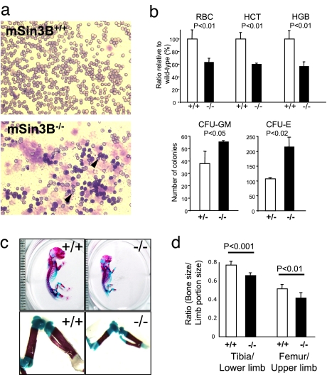
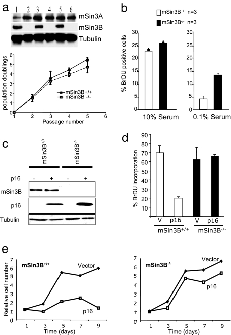
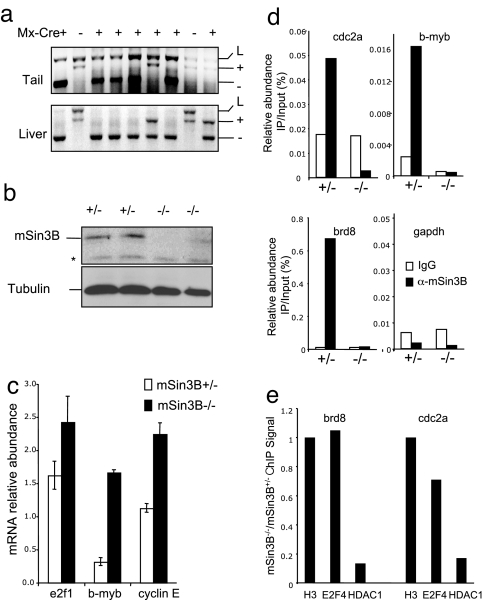
The Sin3-histone deacetylase (HDAC) corepressor complex is conserved from yeast to humans. Mammals possess two highly related Sin3 proteins, mSin3A and mSin3B, which serve as scaffolds tethering HDAC enzymatic activity, and numerous sequence-specific transcription factors to enable local chromatin regulation at specific gene targets. Despite broad overlapping expression of mSin3A and mSin3B, mSin3A is cell-essential and vital for early embryonic development. Here, genetic disruption of mSin3B reveals a very different phenotype characterized by the survival of cultured cells and lethality at late stages of embryonic development with defective differentiation of multiple lineages-phenotypes that are strikingly reminiscent of those associated with loss of retinoblastoma family members or E2F transcriptional repressors. Additionally, we observe that, whereas mSin3B(-/-) cells cycle normally under standard growth conditions, they show an impaired ability to exit the cell cycle with limiting growth factors. Correspondingly, mSin3B interacts physically with the promoters of known E2F target genes, and its deficiency is associated with derepression of these gene targets in vivo. Together, these results reveal a critical role for mSin3B in the control of cell cycle exit and terminal differentiation in mammals and establish contrasting roles for the mSin3 proteins in the growth and development of specific lineages.


Conflict of interest statement

The authors declare no conflict of interest.

Figures

Fig. 1.
Fig. 1.
mSin3B is essential for late embryonic development. (a) (Upper) mSin3B targeted ES cell clones were identified by Southern blot analysis with the probe shown in SI Fig. 5. (Lower) PCR genotyping of embryos harboring the indicated alleles. (b) Western blot on total protein extracts from mSin3BL/+ or mSin3BL/− fibroblasts before (−) or after (+) retroviral-mediated Cre expression using an antibody against mSin3B. Equal amounts of proteins were loaded. *, Cross-reactive band, used as a loading control. (c) Genotype distribution of embryos and offspring from mSin3B heterozygotes intercrosses in a mixed background (E, embryonic day; P, day after birth). *, Found dead. The number of animals analyzed per genotype and time point are indicated in parentheses. (d) Representative litters from mSin3B heterozygote intercrosses, at E14.5 (Upper) or E18.5 (Lower). For each litter, the two embryos on the right are null for mSin3B. (e) Relative weight of mSin3B-null embryos (black bars) compared with their wild-type or heterozygotes littermates (white bars) at E13.5 (Left) or E17.5 (Right). Shown is the average from three litters for each time point.
Fig. 2.
Fig. 2.
mSin3B is required for cellular differentiation during development. (a) Representative smears from peripheral blood of E18.5 mSin3B+/+ (Upper) or mSin3B−/− (Lower) embryos, stained with Wright–Giemsa. Note the high proportion of poorly differentiated erythrocytes in Sin3B-null blood, as evidenced by Howell–Jolly bodies (arrows) and nucleated cells. (b) (Upper) Analysis of peripheral blood defects in Sin3B-null mice. HCT, hematocrit; HGB, hemoglobin. Shown is the average from at least three E18.5 embryos for each genotype. The P values (P) calculated using a t test are indicated. (Lower) In vitro differentiation of E14.5 mSin3B+/− or mSin3B−/− fetal liver cells in CFU-GM or CFU-M. At least three animals were analyzed per genotype. The P values (P) are indicated. (c) Skeletal preparations of E18.5. Sin3B+/+ (Left) and Sin3B−/− (Right) embryos stained with Alcian blue (cartilage) and Alizarin red (bone). Note the diminished ossification of the long bones of the hind limb in the mSin3B−/− embryo. Each graduation corresponds to 1 mm. (d) Quantification of the ratio between bone and limb size in wild-type (white; n = 5) or null (black; n = 6) embryos for Sin3B. The P values are indicated.
Fig. 3.
Fig. 3.
mSin3B is dispensable for cellular proliferation but required for cell cycle exit in vitro. (a) (Upper) Western blot analysis of mSin3B wild-type (lanes 1, 3, 5) and null (lanes 2, 4, 6) primary fibroblasts with the indicated antibodies. (Lower) Growth curves from mSin3B wild-type (n = 3) and null (n = 3) primary fibroblasts at passage 4. (b) BrdU incorporation after a 2-h pulse of 20 μM BrdU by mSin3B wild-type (white bars) or null (black bars) early-passage primary MEFs in 10% serum (Left) or 0.1% serum (Right). At least 200 cells were counted per point. Error bars indicate standard deviations. (c) Western blot of extracts from mSin3B wild-type or null MEFs infected or not with p16 using the indicated antibodies. (d) Proliferation of mSin3B wild-type (n = 3) or null (n = 3) MEFs after p16 or vector (V) infection, assayed by BrdU incorporation. At least 100 cells were counted for each point. (e) Growth curve of mSin3B wild-type (Left) or null (Right) MEFs after p16 or vector infection.
Fig. 4.
Fig. 4.
mSin3B directly represses E2F target genes transcription in vivo. (a) (Upper) PCR for mSin3B alleles on DNA from tail (before pIpC injection) or liver (10 days after pIpC injection) from Sin3B/Mx-Cre transgenic animals. (b) Western blot with the indicated antibodies from liver extracts 10 days after pIpC injection from mSin3BL/+Mx-Cre+ mice (+/−) or mSin3BL/−Mx-Cre+ mice (−/−). *, Nonspecific band. (c) qPCR analysis of mRNA abundance in mSin3B+/− or mSin3B−/− livers as generated in a. Shown is the average abundance of cDNAs after RT-PCR corresponding to the indicated transcripts, normalized to β-2-microglobulin. Three animals for each genotype were used. (d) ChIP assay on the promoter of the indicated E2F target genes and GAPDH, using liver extracts from mSin3BL/+Mx-Cre+ mice (+/−) or mSin3BL/−Mx-Cre+ mice (−/−) with control antibody (IgG, white bars) or anti-mSin3B antibody (black bars). Shown is the average of two independent experiments performed in duplicate. (e) Fold changes of the ChIP signal with E2F4 and HDAC1 antibodies on the promoter of cdc2a and brd8, in liver extracts from mSin3BL/−;Mx-Cre+ mice (−/−) compared with liver extracts from mSin3BL/+;Mx-Cre+ mice (+/−). The results are normalized to the corresponding H3 abundance. Shown is a representative experiment.

References

    1. Knoepfler PS, Eisenman RN. Sin meets NuRD and other tails of repression. Cell. 1999;99:447–450. - PubMed
    1. Silverstein RA, Ekwall K. Sin3: a flexible regulator of global gene expression and genome stability. Curr Genet. 2004;47:1–17. - PubMed
    1. Alland L, et al. Identification of mammalian Sds3 as an integral component of the Sin3/histone deacetylase corepressor complex. Mol Cell Biol. 2002;22:2743–2750. - PMC - PubMed
    1. Kuzmichev A, Zhang Y, Erdjument-Bromage H, Tempst P, Reinberg D. Role of the Sin3-histone deacetylase complex in growth regulation by the candidate tumor suppressor p33(ING1). Mol Cell Biol. 2002;22:835–848. - PMC - PubMed
    1. Laherty CD, et al. SAP30, a component of the mSin3 corepressor complex involved in N-CoR-mediated repression by specific transcription factors. Mol Cell. 1998;2:33–42. - PubMed

Publication types

LinkOut - more resources