. 2008 May;154(1):4-12.
doi: 10.1038/bjp.2008.82. Epub 2008 Mar 10.

Photodynamic activity of BAM-SiPc, an unsymmetrical bisamino silicon(IV) phthalocyanine, in tumour-bearing nude mice

Affiliations

Photodynamic activity of BAM-SiPc, an unsymmetrical bisamino silicon(IV) phthalocyanine, in tumour-bearing nude mice

S C H Leung et al. Br J Pharmacol. 2008 May.

Abstract

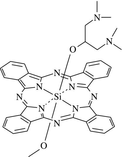
Background and purpose: Ever since the discovery of photodynamic therapy, there has been a continuous search for more potent photosensitizers. Towards that end, we have synthesized a number of novel phthalocyanine derivatives. The unsymmetrical bisamino silicon(IV) phthalocyanine BAM-SiPc is one of the most potent compounds. In in vitro cell culture, it exhibits high phototoxicity against a number of cancer cell lines.

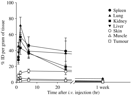
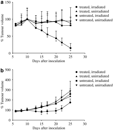
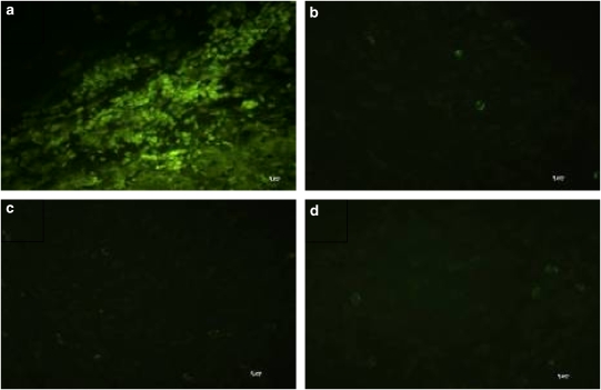
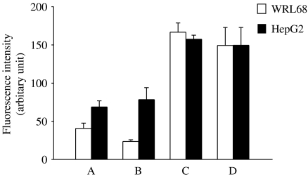
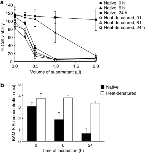
Experimental approach: In the present investigation, the in vivo effect of BAM-SiPc was studied in the tumour-bearing nude mice model. The biodistribution of BAM-SiPc was followed to evaluate its tumour selectivity and rate of clearance. The tumour volume in the hepatocarcinoma HepG2- and the colorectal adenocarcinoma HT29-bearing nude mice was measured after photodynamic therapy. The level of intrinsic toxicity induced was also investigated. Finally, the metabolism of BAM-SiPc in the 'normal' WRL68 liver cells and the hepatocarcinoma HepG2 cells was compared.

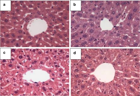
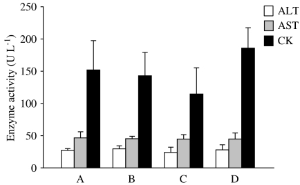
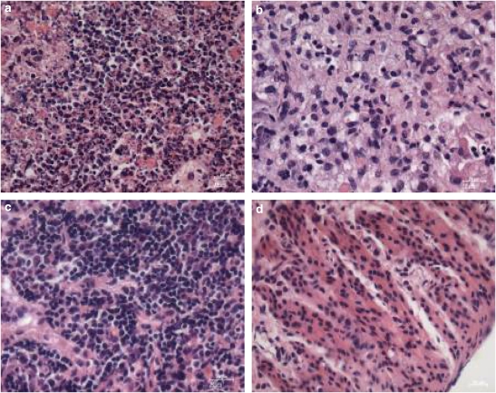
Key results: The results not only showed significant tumour regression of HepG2 and growth inhibition of HT29 in the tumour-bearing nude mice, but also no apparent hepatic or cardiac injury with the protocol used. Histological analyses showed that apoptosis was induced in the solid tumour. BAM-SiPc could be metabolized by WRL68 liver cells but not by the hepatocarcinoma HepG2 cells. Unfortunately, BAM-SiPc did not show any specific targeting towards the tumour tissue.

Conclusions and implications: The efficiency of BAM-SiPc in inhibiting tumour growth makes it a good candidate for further evaluation. Enhancement of its uptake in tumour tissue by conjugation with biomolecules is currently under investigation.


Figures

Figure 1
Figure 1
Chemical structure of BAM-SiPc.
Figure 2
Figure 2
Tissue distribution of BAM-SiPc. One μmol of BAM-SiPc kg−1 body weight was injected, intravenously, into the HepG2-bearing nude mice. Mice were killed at different timed intervals. Tissues were excised and homogenized with DMF. The DMF fraction was subjected to fluorescence measurement to determine the amount of BAM-SiPc, expressed as % initial dose g−1 tissue. Data are expressed as mean±s.d., n=5.
Figure 3
Figure 3
Photodynamic therapy of (a) HepG2- and (b) HT29- bearing nude mice treated with BAM-SiPc. In vivo photodynamic treatment was started on day 9 after inoculation of tumour cells. One μmol BAM-SiPc kg−1 body weight was injected, intravenously, into the tumour-bearing nude mice. Twenty-four hours after the injection, the mice were anaesthetized and irradiated with 30 J cm−2. Data represent mean±s.d., n=8 and n=6 for each group of HepG2- and HT29-bearing mice respectively. The tumour volume of the mice on day 10 was taken as 100%. *P<0.05, when the PDT (treated and irradiated) group was compared with all the three control groups.
Figure 4
Figure 4
Typical microscopy images obtained after H & E staining of liver sections from PDT-treated mice, showing the region around the hepatic artery. Fifteen days after PDT, livers were excised from the nude mice and sections were stained. Nuclei were stained by haematoxylin solution (purple) and the cytoplasm was counterstained by eosin solution (pink). (a) PDT-treated; (b) treated but not irradiated; (c) untreated but irradiated and (d) untreated and not irradiated. Original magnification is × 40.
Figure 5
Figure 5
Plasma enzyme activities of the PDT-treated mice. At 15 days after PDT, plasma samples were obtained from different groups of mice for determination of enzyme activity. (a) PDT-treated; (b) treated but not irradiated; (c) untreated but irradiated and (d) untreated and not irradiated. Data represent mean±s.d., n=6.
Figure 6
Figure 6
Typical fluorescence microscopy images obtained after TUNEL staining of solid tumour from PDT-treated mice. At 24 h after PDT, the HepG2 solid tumours were excised from the mice and the histological sections were stained by the TUNEL reaction mixture. (a) PDT-treated; (b) treated but not irradiated; (c) untreated but irradiated and (d) untreated and not irradiated. Original magnification is × 40.
Figure 7
Figure 7
Typical microscopy images obtained after H & E staining of solid tumour from PDT-treated mice. At 15 days after PDT, the HepG2 solid tumours were excised from the nude mice and sections were stained. (a) PDT-treated; (b) treated but not irradiated; (c) untreated but irradiated and (d) untreated and not irradiated. Original magnification is × 40.
Figure 8
Figure 8
Comparison of BAM-SiPc retention in WRL68 and HepG2 cells. Cells were incubated with 0.5 μM BAM-SiPc for 2 h for uptake before the unbound photosensitizer was removed by washing. The cells were then lysed with DMF either (a) immediately, or (b) after 24 h of incubation with the culture medium. The residual BAM-SiPc was estimated by fluorescence measurement. Two controls were performed: (c) cells lysed with DMF before adding BAM-SiPc and (d) BAM-SiPc, cell-free control. Data represent mean±s.d., n=3.
Figure 9
Figure 9
Effect of pre-incubation with mouse liver homogenate on the photocytotoxicity of BAM-SiPc. (a) BAM-SiPc (8 μM) was first incubated with native or heat-denatured homogenate for different times. After centrifugation, aliquots of the supernatant were tested for in vitro photodynamic activity by MTT assay using the HepG2 cells. Data represent mean±s.e.mean from three independent experiments, each performed in triplicate. (b) The residual BAM-SiPc after incubation with native or heat-denatured liver homogenate was estimated by absorbance measurement at 674 nm. Data represent mean±s.d., n=3.

Comment in

References

    1. Bellnier DA, Greco WR, Loewen GM, Nava H, Oseroff AR, Pandey RK, et al. Population pharmacokinetics of the photodynamic therapy agent 2-[1-hexyloxyethyl]-2-devinyl pyropheophorbide-a in cancer patients. Cancer Res. 2003;63:1806–1813. - PubMed
    1. Bourdon O, Laville I, Carrez D, Croisy A, Fedel P, Kasselouri A, et al. Biodistribution of meta-tetra(hydroxyphenyl)chlorin incorporated into surface-modified nanocapsules in tumor-bearing mice. Photochem Photobiol Sci. 2002;1:709–714. - PubMed
    1. Brasseur N, Nguyen TL, Langlois R, Ouellet R, Marengo S, Houde D, et al. Synthesis and photodynamic activities of silicon 2, 3- naphthalocyanine derivatives. J Med Chem. 1994;37:415–420. - PubMed
    1. Brasseur N, Ouellet R, La Madeleine C, van Lier JE. Water-soluble aluminium phthalocyanine-polymer conjugates for PDT: photodynamic activities and pharmacokinetics in tumour-bearing mice. Br J Cancer. 1999;80:1533–1541. - PMC - PubMed
    1. Brown SB, Brown EA, Walker I. The present and future role of photodynamic therapy in cancer treatment. Lancet Oncol. 2004;5:497–508. - PubMed

Publication types

MeSH terms