

Three cases of small hepatocellular carcinoma presenting as obstructive jaundice
- PMID: 18333040
- PMCID: PMC2020647
- DOI: 10.1080/13651820310017129
Three cases of small hepatocellular carcinoma presenting as obstructive jaundice
Abstract
Background: Despite improved diagnostic tools, it is often difficult to make a correct diagnosis of small hepatocellular carcinoma (HCC) in patients with obstructive jaundice.
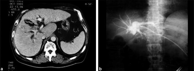
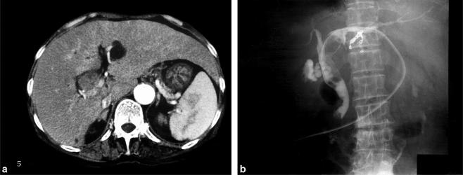
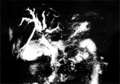
Case outlines: Three cases of small HCC (<2 cm diameter) presenting as obstructive jaundice are reported. All tumours were initially diagnosed as hilar cholangiocarcinoma based on ultrasonography, computed tomography, cholangiography and angiography. Because of insufficient hepatic function, none of the patients underwent hepatic resection. One patient died 8 months after first admission to our hospital, another died of disseminated intravascular coagulation I month after admission, and the third was treated with hepatic arterial infusion chemotherapy and survived >36 months.
Conclusion: It is important to consider HCC in the diagnosis of obstructive jaundice in patients who are predisposed to HCC because of liver cirrhosis and/or chronic viral hepatitis, and have elevated serum alpha-fetoprotein.
Figures
References
-
- Lin TY, Chen KM, Chen YR, Lin WS, Wang TH, Sung JL. Icteric type hepatoma. Med Chir Dig. 1975;4:267–70. - PubMed
-
- Lau WY, Leung JW, Li AK. Management of hepatocellular carcinoma presenting as obstructive jaundice. Am J Surg. 1990;160:280–2. - PubMed
-
- Huang GT, Sheu JC, Lee HS, Lai MY, Wang TH, Chen DS. Icteric type hepatocellular carcinoma: revisited 20 years later. J Gastroenterol. 1998;33:53–6. - PubMed
LinkOut - more resources
Full Text Sources
