. 2008 Mar;16(3):371-9.
doi: 10.1016/j.str.2007.12.023.

Colicin N binds to the periphery of its receptor and translocator, outer membrane protein F

Affiliations

Colicin N binds to the periphery of its receptor and translocator, outer membrane protein F

Thomas G Baboolal et al. Structure. 2008 Mar.

Abstract

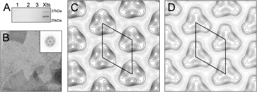
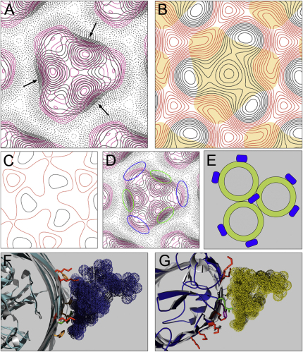
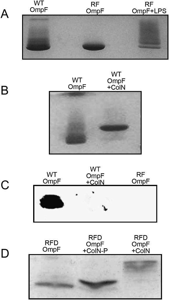
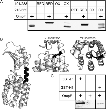
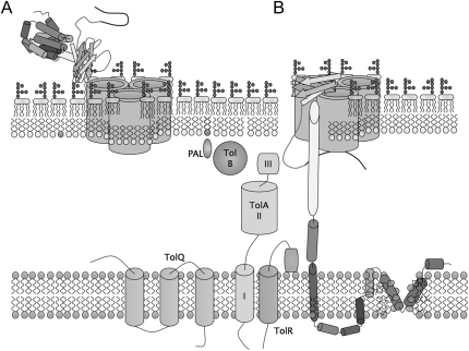
Colicins kill Escherichia coli after translocation across the outer membrane. Colicin N displays an unusually simple translocation pathway, using the outer membrane protein F (OmpF) as both receptor and translocator. Studies of this binary complex may therefore reveal a significant component of the translocation pathway. Here we show that, in 2D crystals, colicin is found outside the porin trimer, suggesting that translocation may occur at the protein-lipid interface. The major lipid of the outer leaflet interface is lipopolysaccharide (LPS). It is further shown that colicin N binding displaces OmpF-bound LPS. The N-terminal helix of the pore-forming domain, which is not required for pore formation, rearranges and binds to OmpF. Colicin N also binds artificial OmpF dimers, indicating that trimeric symmetry plays no part in the interaction. The data indicate that colicin is closely associated with the OmpF-lipid interface, providing evidence that this peripheral pathway may play a role in colicin transmembrane transport.


Figures

Figure 1
Figure 1
2D Crystals of OmpF + Colicin N-RP Are Visibly Different to OmpF Alone (A) Coomassie-stained SDS-PAGE of OmpF/colN-RP 2D-crystal (LPR, 1:2 w/w) together with several wash samples. (B) An electron micrograph showing an area of negatively stained OmpF/colN-RP 2D-crystal. Scale bar = 100 nm. The insert shows the relevant diffraction pattern. (C) Projection map showing the density derived from four merged OmpF/colN-RP crystals. The unit cell is indicated by the solid line. (D) Projection map showing the density derived from four merged OmpF 2D-crystals. The unit cell is indicated by the solid line.
Figure 2
Figure 2
OmpF/ColN-RP Crystals Show Increased Peripheral Density at Monomer-Monomer Interfaces but Reduced Density where LPS Binds (A) Superposition of the merged and scaled projection maps from Figure 1 (OmpF crystal in magenta, OmpF/colN-RP crystal in black). The arrows indicate the areas of extra density contributed to the crystal by the presence of colN-RP. (B) A superposition of the OmpF footprint (solid orange) with difference map showing density due to colN-RP within the OmpF/colN-RP crystals. The colN-RP projection map was calculated from the subtraction of the merged and scaled OmpF data from that of the OmpF/colN-RP data in Fourier space. Negative contours are shown in red with positive contours shown in black. (C) A difference map showing the subtraction of two independently merged OmpF maps. Contours are at the same scale and orientation as in (B). (D) Superposition of the merged and scaled projection maps as in (A), with the areas of extra density in OmpF crystal indicated with blue and those of the complex in green. (E) A schematic of OmpF with bound LPS in those positions predicted by the work of Hoenger et al. (1990). The central LPS molecule on the trimeric axis of symmetry is not supported by more recent X-ray data, because no suitable cavity exists (Cowan et al., 1992). (F) FhuA with bound LPS (PDB code: 1QFG) (Ferguson et al., 2000). Indicated are those residues thought to constitute an LPS-binding motif (Lys in red, Arg in green, and Phe of the hydrophobic boundary in orange). (G) A proposed LPS-binding site located around Arg 235 based on the work of Ferguson et al. (2000) (Lys in red, Arg in green, Tyr in white, and Trp in purple; also see the Supplemental Data).
Figure 3
Figure 3
LPS Is Displaced from OmpF by Colicin N Complex Formation (A) The effect of LPS on the elecrophoretic migration of OmpF (WT OmpF, RF OmpF, and RF OmpF+LPS). (B) The effect of colN-RP on the electrophoretic migration of OmpF showing the shift in migration of OmpF owing to the increase mass of the complex and the loss of OmpF bound LPS. (C) Western blot using WN1 222-5 antibody (Di Padova et al., 1993) after SDS-PAGE to detect LPS. LPS is bound to OmpF but largely removed by colicin N addition, and the antibody shows no nonspecific binding to refolded LPS-free OmpF. (D) The effect of full-length colicin N (+ColN) and colicin P-domain (+ColN-P) on the electrophoretic migration of RFD (refolded dimeric) OmpF. The increase in mass of the RFD OmpF band is due to the increased molecular weight of the complex.
Figure 4
Figure 4
Complex Formation Involves Helix-1 of the Pore-Forming Domain (A) Coomassie-stained SDS-PAGE showing binding of the reduced (RED) and oxidized (OX) forms of colN N191C-A288C and colN Y213C-V352C to trimeric OmpF. Each disulfide fixes one end of Helix-1 in the native conformation. OmpF alone occurs as a doublet caused by LPS. Colicin/OmpF complex migrates at a higher MW, formation of which is inhibited by disulfide bond formation (OX). (B) Structure of colicin N (PDB code: 1A87) with the two disulfide bridges used in panel A represented with arrows and zoomed views. Dark region indicates the region of helix-1 fused to GST in GST-H1. (C) Anti-GST western blot showing the binding of OmpF to fusions of GST to the entire pore-forming domain GST-P or the first helix of the pore domain (GST-H1). GST-H1 was easily proteolyzed, causing the low intensity.
Figure 5
Figure 5
Possible Arrangement and Translocation Mechanism for Colicin N (A) A schematic representation of initial interaction of the colicin N receptor binding domain with OmpF in the E. coli outer membrane. (B) The suggested arrangement of unfolded colicin N according to data from this study. The pore-forming domain unfolds and interacts with the external surface of OmpF, filling the cleft between two monomers to agree with EM density while displacing LPS. The unfolded pore-forming domain is sufficient to make the ion channel, whereas the suggested rearrangement of helix-1 would be prevented by the disulfide bonds that prevented complex formation.

References

    1. Anderluh G., Dalla Serra M., Viero G., Guella G., Macek P., Menestrina G. Pore formation by equinatoxin II, a eukaryotic protein toxin, occurs by induction of nonlamellar lipid structures. J. Biol. Chem. 2003;278:45216–45223. - PubMed
    1. Bainbridge G., Armstrong G.A., Dover L.G., Whelan K.F., Lakey J.H. Displacement of OmpF loop 3 is not required for the membrane translocation of colicins N and A in vivo. FEBS Lett. 1998;432:117–122. - PubMed
    1. Barlic A., Gutierrez-Aguirre I., Caaveiro J.M.M., Cruz A., Ruiz-Arguello M.B., Perez-Gil J., Gonzalez-Manas J.M. Lipid phase coexistence favors membrane insertion of equinatoxin-II, a pore-forming toxin from Actinia equina. J. Biol. Chem. 2004;279:34209–34216. - PubMed
    1. Baty D., Lakey J., Pattus F., Lazdunski C. A 136-amino-acid-residue COOH-terminal fragment of colicin A is endowed with ionophoric activity. Eur. J. Biochem. 1990;189:409–413. - PubMed
    1. Benedetti H., Lloubes R., Lazdunski C., Letellier L. Colicin A unfolds during its translocation in Escherichia coli cells and spans the whole cell envelope when its pore has formed. EMBO J. 1992;11:441–447. - PMC - PubMed

Publication types

MeSH terms

LinkOut - more resources