

Mechanism of activation and inhibition of the HER4/ErbB4 kinase
- PMID: 18334220
- PMCID: PMC2858219
- DOI: 10.1016/j.str.2007.12.016
Mechanism of activation and inhibition of the HER4/ErbB4 kinase
Abstract
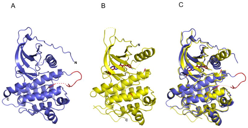
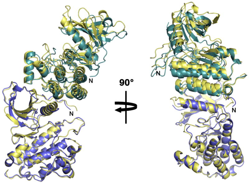
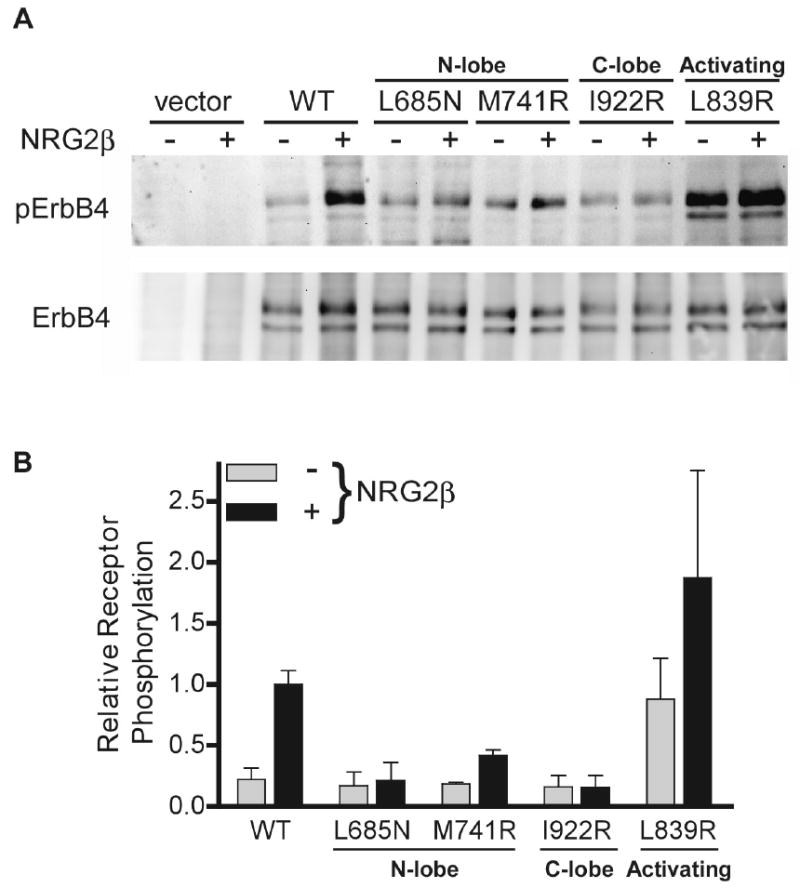
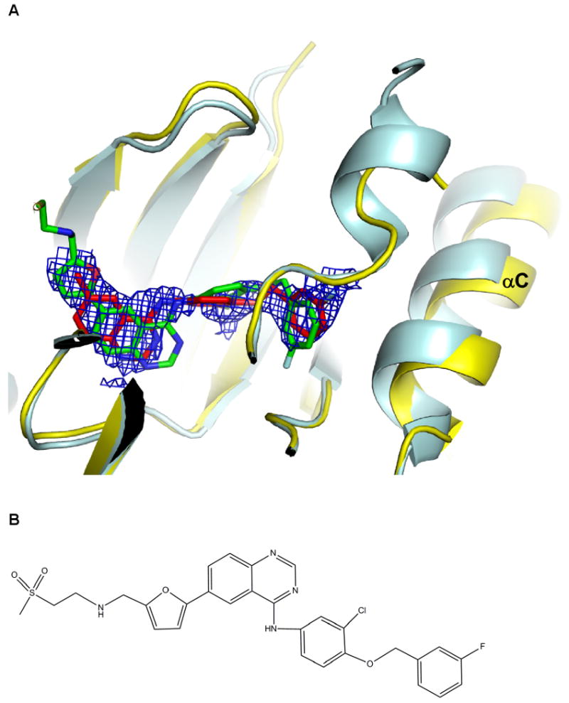
HER4/ErbB4 is a ubiquitously expressed member of the EGF/ErbB family of receptor tyrosine kinases that is essential for normal development of the heart, nervous system, and mammary gland. We report here crystal structures of the ErbB4 kinase domain in active and lapatinib-inhibited forms. Active ErbB4 kinase adopts an asymmetric dimer conformation essentially identical to that observed to be important for activation of the EGF receptor/ErbB1 kinase. Mutagenesis studies of intact ErbB4 in Ba/F3 cells confirm the importance of this asymmetric dimer for activation of intact ErbB4. Lapatinib binds to an inactive form of the ErbB4 kinase in a mode equivalent to its interaction with the EGF receptor. All ErbB4 residues contacted by lapatinib are conserved in the EGF receptor and HER2/ErbB2, which lapatinib also targets. These results demonstrate that key elements of kinase activation and inhibition are conserved among ErbB family members.
Figures
Comment in
-
A trigger squeezed.Structure. 2008 Mar;16(3):332-4. doi: 10.1016/j.str.2008.02.002. Structure. 2008. PMID: 18334205 No abstract available.
References
-
- Berger MB, Mendrola JM, Lemmon MA. ErbB3/HER3 does not homodimerize upon neuregulin binding at the cell surface. FEBS Lett. 2004;569:332–336. - PubMed
-
- Brignola PS, Lackey K, Kadwell SH, Hoffman C, Horne E, Carter HL, Stuart JD, Blackburn K, Moyer MB, Alligood KJ, et al. Comparison of the biochemical and kinetic properties of the type 1 receptor tyrosine kinase intracellular domains. Demonstration of differential sensitivity to kinase inhibitors. J Biol Chem. 2002;277:1576–1585. - PubMed
-
- Brunger AT, Adams PD, Clore GM, DeLano WL, Gros P, Grosse-Kunstleve RW, Jiang JS, Kuszewski J, Nilges M, Pannu NS, et al. Crystallography & NMR system: A new software suite for macromolecular structure determination. Acta Crystallogr D Biol Crystallogr. 1998;54:905–921. - PubMed
Publication types
MeSH terms
Substances
Associated data
- Actions
- Actions
- Actions
Grants and funding
LinkOut - more resources
Full Text Sources
Other Literature Sources
Molecular Biology Databases
Research Materials
Miscellaneous
