. 2008 May;36(8):2608-18.
doi: 10.1093/nar/gkn104. Epub 2008 Mar 11.

Sequence homology and microhomology dominate chromosomal double-strand break repair in African trypanosomes

Affiliations

Sequence homology and microhomology dominate chromosomal double-strand break repair in African trypanosomes

Lucy Glover et al. Nucleic Acids Res. 2008 May.

Abstract

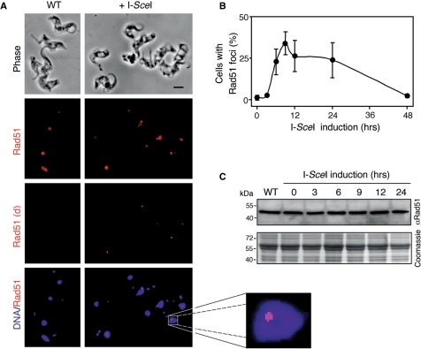
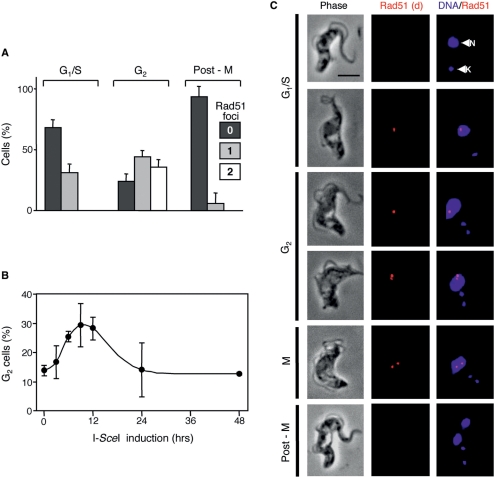
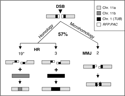
Genetic diversity in fungi and mammals is generated through mitotic double-strand break-repair (DSBR), typically involving homologous recombination (HR) or non-homologous end joining (NHEJ). Microhomology-mediated joining appears to serve a subsidiary function. The African trypanosome, a divergent protozoan parasite, relies upon rearrangement of subtelomeric variant surface glycoprotein (VSG) genes to achieve antigenic variation. Evidence suggests an absence of NHEJ but chromosomal repair remains largely unexplored. We used a system based on I-SceI meganuclease and monitored temporally constrained DSBR at a specific chromosomal site in bloodstream form Trypanosoma brucei. In response to the lesion, adjacent single-stranded DNA was generated; the homologous strand-exchange factor, Rad51, accumulated into foci; a G(2)M checkpoint was activated and >50% of cells displayed successful repair. Quantitative analysis of DSBR pathways employed indicated that inter-chromosomal HR dominated. HR displayed a strong preference for the allelic template but also the capacity to interact with homologous sequence on heterologous chromosomes. Intra-chromosomal joining was predominantly, and possibly exclusively, microhomology mediated, a situation unique among organisms examined to date. These DSBR pathways available to T. brucei likely underlie patterns of antigenic variation and the evolution of the vast VSG gene family.


Figures

Figure 1.
Figure 1.
Experimental system to study DSBR. (A) The schematic illustrates Tb11.02.2110/2120 loci on chromosome 11a and b (WT: top) and the same loci after insertion of the RsP cassette (RsP: bottom). The arrowhead in the top panel indicates the RsP insertion site, 209-bp downstream of the 2110 stop codon. Bars above the maps indicate the locations of the 2110 and RFP probes. The sizes of the HindIII fragments expected on Southern blots are indicated below the maps. Black boxes indicate tubulin (TUB) sequences flanking the RsP cassette. H, HindIII sites. (B) Southern blot analysis indicated RsP insertion on chromosome 11a. DNA was digested with HindIII. See fragment sizes in (A). (C) Schematic illustration of possible DSBR mechanisms. HR requires extensive homologous sequence; MMJ requires only short stretches and NHEJ requires little or none. HR can use templates from chromosome 11b (top) or from TUB genes on chromosome 1 (bottom). Portions of RFP and PAC would be expected to be retained following NHEJ or MMJ.
Figure 2.
Figure 2.
Physical monitoring of DNA resection and repair. (A) Monitoring ssDNA adjacent to the lesion by slot-blot analysis. Genomic DNA samples were extracted at various times following I-SceI-induction. Ninety percent of each sample was ‘native’ (n) and the remainder denatured (d). The probes used on each blot are indicated on the right. 7240 is a distal, chromosome 11 control. The schematic map indicates the location of the probes (black) in relation to the lesion (DSB). (B) Kinetics of ssDNA formation. Phoshorimager analysis was used to quantify the signals in (A). (C) Monitoring repair by Southern blot analysis. Genomic DNA extracted at various times following I-SceI-induction was digested with HindIII and subjected to Southern blot analysis using the probes indicated. Arrowheads indicate the fragments expected following HR between chromosome 11a and 11b or the TUB locus on chromosome 1 (see Figure 1). For chromosome 11, 7240 served as a loading control. The schematic illustrates dominant allelic HR with chromosome 11b. (D) Kinetics of repair by HR with chromosome 11b. Phoshorimager analysis was used to quantify the signals in (C).
Figure 5.
Figure 5.
Survivors display repair via homologous recombination and microhomology-mediated joining. (A) Survivorship. To generate mostly clonal populations, we distributed ∼96 cells over 288 wells (three 96-well plates). This yielded 93 (no Tet) and 53 (+Tet) wells, respectively with live cells detected after 1 week of growth. (B) Genomic DNA was extracted from survivors of I-SceI-induction, digested with HindIII and subjected to Southern blot analysis using the probes indicated (see Figure 2C for other details). In the αTUB panel, the intense band in every sample represents the tandem array on chromosome 1; the bands in the un-induced (U) and all survivor lanes represent the TetR insertion at the TUB locus and the additional 9.5-kb band in track 9 (arrowhead) confirmed ectopic recombination with chromosome 1. (C) Joined junctions. The PCR, end-joining survey employed primers at the extreme ends of the RFP/PAC ORF and DNA from a large population of cells after 7 days of Tet exposure (Tet+). DNA from cells prior to Tet exposure (Tet) served as a control. Junction sequences from two major PCR products (arrowheads 1 and 2) and from four independent cloned survivors are shown on the right. The RFP (top row), PAC (bottom row) and junction sequences (middle row) are aligned with microhomology highlighted. The number of nucleotides deleted in each case and the number of clones representing each sequence are indicated.
Figure 3.
Figure 3.
Rad51 accumulates at sub-nuclear foci in response to DSBs. (A) Immunofluorescence analysis of Rad51 in wild-type (WT) cells and in Sce2110 cells 9 h after I-SceI-induction. Rad51 signals are shown before and after deconvolution (d). DNA was counter-stained with DAPI. Scale bar, 5 μm. An expanded view of a nucleus with a prominent Rad51 focus is shown to the right. (B) Rad51 foci kinetics. The proportion of nuclei with Rad51 foci were counted at different times after I-SceI-induction. n = 200 at each time point. Error bars, SD. (C) Rad51 levels remain constant during DSBR. Western blotting with anti-Rad51 and a series of protein samples extracted at different times after I-SceI-induction. An equivalent Coomassie-stained gel served as a loading-control. The predicted Mwt of TbRad51 is 41 kDa.
Figure 4.
Figure 4.
Rad51 foci and the G2M cell cycle checkpoint. Cells were processed for Rad51 immunofluorescence microscopy and DNA was counter-stained with DAPI. (A) The bar-chart shows the proportion of Sce2110 cells at different phases of the cell cycle with zero, one or two Rad51 foci 12 h after I-SceI-induction. Cell cycle phase was defined by the number of nuclei (N) and kinetoplasts (K) as determined by DAPI staining. n = 50 at each cell cycle phase. Error bars, SD. (B) G2M phase (1N2K) kinetics. The proportion of 1N2K cells was counted at different times after I-SceI-induction. n = 200 at each time point. Error bars, SD. (C) Immunofluorescence analysis of Rad51 in Sce2110 cells after I-SceI-induction. Rad51 signals are shown after deconvolution (d). N, nucleus; K, kinetoplast. Scale bar, 5 μm.
Figure 6.
Figure 6.
Summary of double-strand break-repair mechanisms in T. brucei. Approximately 57% of cells recover from a double-strand break (DSB) on chromosome 11a. Three distinct modes of repair were detected among at least 26 independent repair events. Resection is illustrated and the numbers of events detected are shown in each case. Asterisk denotes ∼85% of repair occurs via allelic recombination with chromosome 11b (see the text).

References

    1. Simpson AG, Stevens JR, Lukes J. The evolution and diversity of kinetoplastid flagellates. Trends Parasitol. 2006;22:168–174. - PubMed
    1. Horn D. The molecular control of antigenic variation in Trypanosoma brucei. Curr. Mol. Med. 2004;4:563–576. - PubMed
    1. Morrison LJ, Majiwa P, Read AF, Barry JD. Probabilistic order in antigenic variation of Trypanosoma brucei. Int. J. Parasitol. 2005;35:961–972. - PubMed
    1. Robinson NP, Burman N, Melville SE, Barry JD. Predominance of duplicative VSG gene conversion in antigenic variation in African trypanosomes. Mol. Cell Biol. 1999;19:5839–5846. - PMC - PubMed
    1. Callejas S, Leech V, Reitter C, Melville S. Hemizygous subtelomeres of an African trypanosome chromosome may account for over 75% of chromosome length. Genome Res. 2006;16:1109–1118. - PMC - PubMed

Publication types