. 2008 Jun 15;17(12):1855-66.
doi: 10.1093/hmg/ddn081. Epub 2008 Mar 11.

Calpain 3 is a modulator of the dysferlin protein complex in skeletal muscle

Affiliations

Calpain 3 is a modulator of the dysferlin protein complex in skeletal muscle

Yanchao Huang et al. Hum Mol Genet. .

Abstract

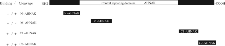
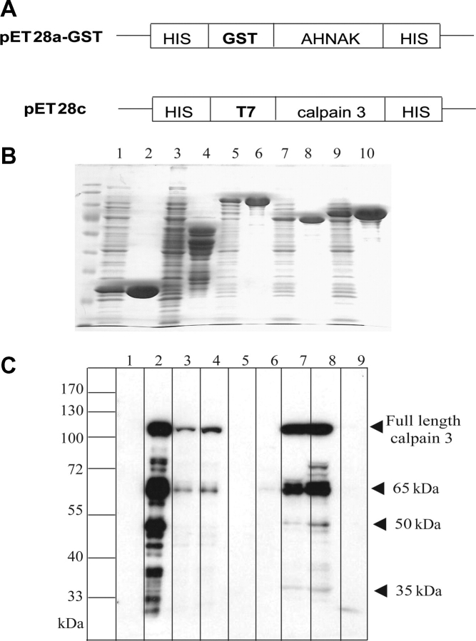
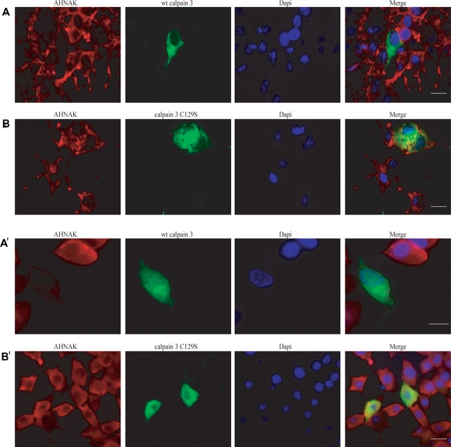
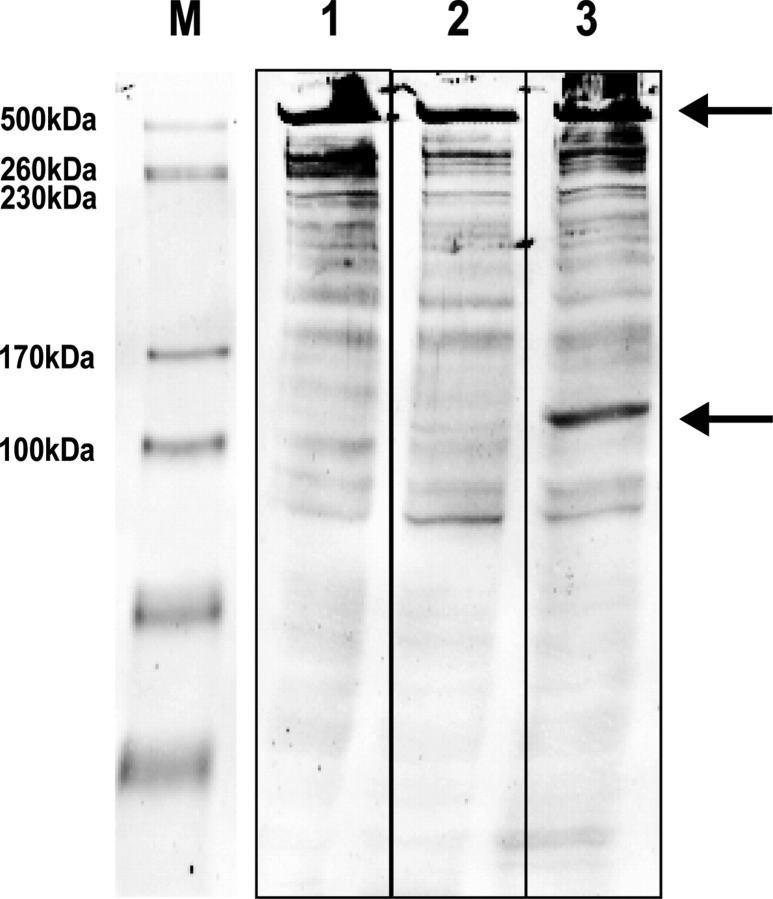
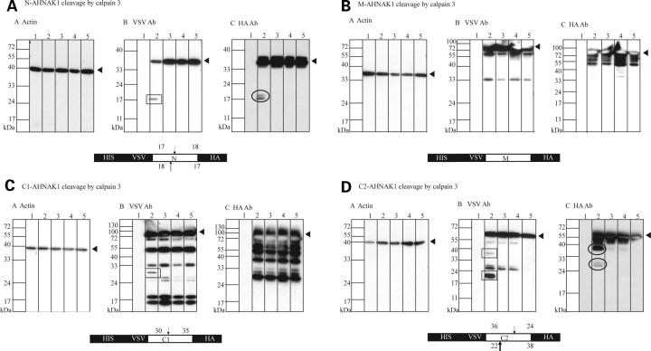
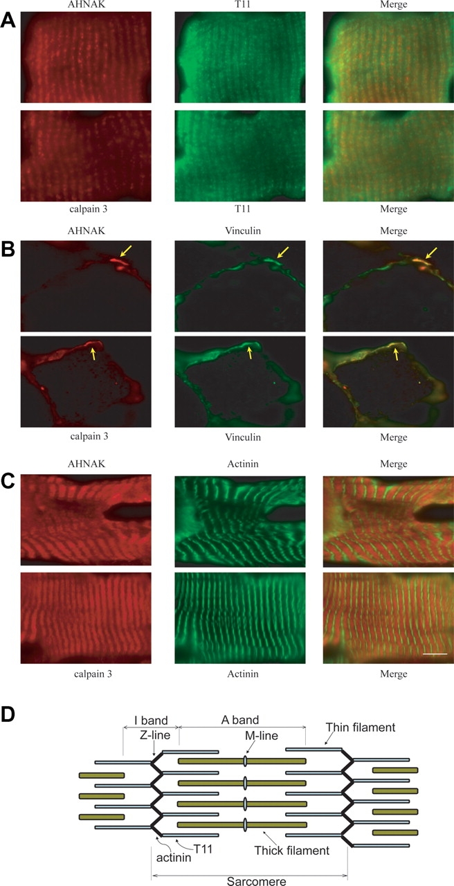
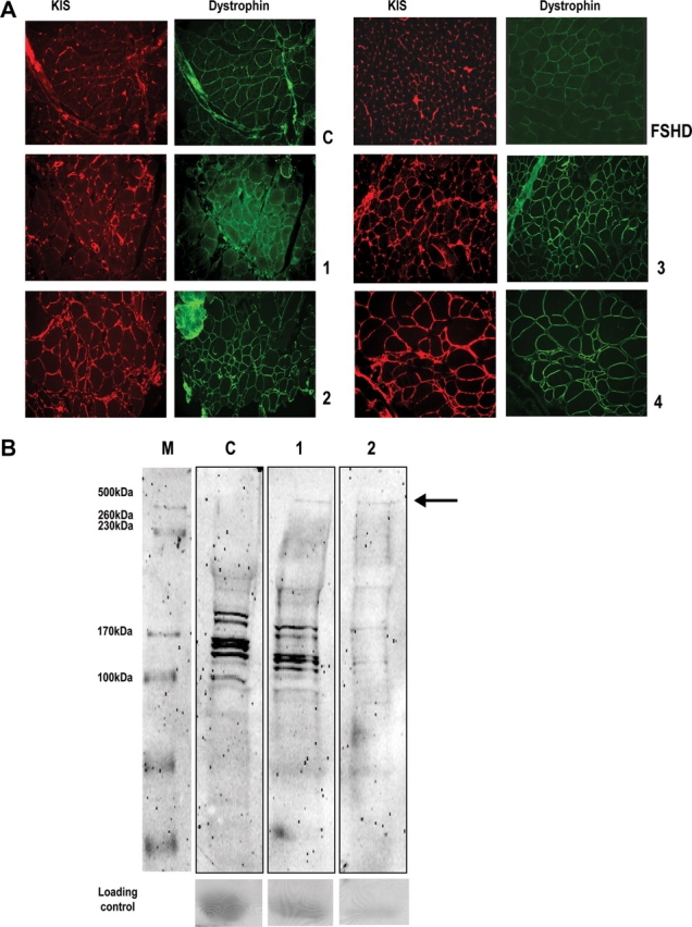
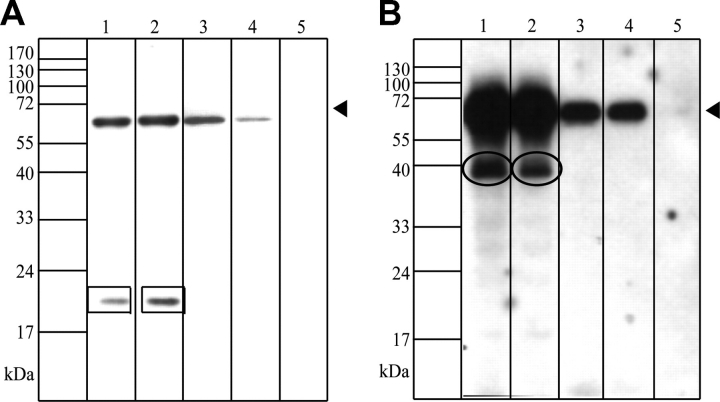
Muscular dystrophies comprise a genetically heterogeneous group of degenerative muscle disorders characterized by progressive muscle wasting and weakness. Two forms of limb-girdle muscular dystrophy, 2A and 2B, are caused by mutations in calpain 3 (CAPN3) and dysferlin (DYSF), respectively. While CAPN3 may be involved in sarcomere remodeling, DYSF is proposed to play a role in membrane repair. The coexistence of CAPN3 and AHNAK, a protein involved in subsarcolemmal cytoarchitecture and membrane repair, in the dysferlin protein complex and the presence of proteolytic cleavage fragments of AHNAK in skeletal muscle led us to investigate whether AHNAK can act as substrate for CAPN3. We here demonstrate that AHNAK is cleaved by CAPN3 and show that AHNAK is lost in cells expressing active CAPN3. Conversely, AHNAK accumulates when calpain 3 is defective in skeletal muscle of calpainopathy patients. Moreover, we demonstrate that AHNAK fragments cleaved by CAPN3 have lost their affinity for dysferlin. Thus, our findings suggest interconnectivity between both diseases by revealing a novel physiological role for CAPN3 in regulating the dysferlin protein complex.


Figures

Figure 1.
Figure 1.
Schematic representation of full length AHNAK and the different constructs used in these studies. On the left, the ability to bind and/or act as a substrate for calpain 3 is indicated for each AHNAK fragment.
Figure 2.
Figure 2.
Identification of the interaction sites between AHNAK and inactive calpain 3 C129S. (A) Scheme of vectors for expressing AHNAK fusion proteins and calpain 3 C129S fusion protein. (B) Induced GST (lane 1) or GST bound to the affinity beads (lane 2); lanes 3–10 represent induced and affinity beads-bound GST-N-AHNAK (lanes 3–4), GST-M-AHNAK (lanes 5–6), GST-C1-AHNAK (lanes 7–8) and GST-C2-AHNAK (lanes 9–10), respectively. Equal amounts of GST fusion proteins representing different domains of AHNAK or GST were used in pull-down assays with T7-tagged calpain 3 C129S fusion protein as described in Materials and Methods. Bound proteins were separated by SDS–PAGE and analyzed by immunoblotting with anti-T7 HRP. (C) Lanes 1–4 represent uninduced-, induced-, soluble-, and precleared T7-tagged calpain 3 C129S, respectively. Lanes 5–8 represent GST-N-AHNAK, GST-M-AHNAK, GST-C1-AHNAK and GST-C2-AHNAK pull-down fractions while lane 9 shows the result with GST as negative control. As shown, GST-C1-AHNAK and GST-C2-AHNAK pulled down T7-tagged full length calpain 3 C129S fusion protein and its degradation products (arrowheads). GST-N-AHNAK, GST-M-AHNAK and GST did not interact with calpain 3 C129S fusion proteins. A molecular mass marker is indicated on the left.
Figure 3.
Figure 3.
Cleavage of AHNAK by calpain 3 downregulates AHNAK in cell culture. To evaluate the function of AHNAK cleavage by calpain 3 in COS-1 (A and B) and 3T3 cells (A' and B'), we immunocytochemically examined wt calpain 3 and calpain 3 C129S expressing cells using KIS antibody to detect AHNAK. Absence of endogenous AHNAK was observed in wt calpain 3 overexpressing cells (A and A'), compared with non-transfected cells. In contrast, normal AHNAK signals were observed in calpain 3 C129S expressing cells (B and B'). Bars represent 20 µm.
Figure 4.
Figure 4.
Cleavage of endogenous AHNAK by recombinant calpain 3. Cos cells were transfected with either mock (lane 1), inactive calpain 3-GFP (C129S mutant) (lane 2) or active calpain 3-GFP (lane 3). Forty-eight hours after transfection cells were harvested and analyzed on western blot with a KIS/CQL antibody mixture. The arrows point out full length AHNAK and a 120 kDa band which arises upon active calpain 3 expression.
Figure 5.
Figure 5.
Specific domains of AHNAK are cleaved by calpain 3 in cell culture. Four AHNAK fusion proteins representing N- (A), M- (B), C1- (C) and C2- of AHNAK (D), respectively, were expressed in the presence of active or inactive calpain 3 C129S to assess whether AHNAK can act as a substrate for calpain 3. The AHNAK fusion proteins were N-terminally tagged with a VSV tag and C-terminally tagged with a HA tag to detect N-terminal and C-terminal AHNAK cleavage products. Actin detection was used for equal loading control (a). Antibodies recognizing the N-terminal VSV tag (b) and C-terminal HA tag (c) were used to detect proteolytic cleavage products on western blots. In all panels, lane 1–5 represent lysates of non-transfected COS-1 cells, co-transfection of AHNAK fusion construct with calpain 3, co-transfection of AHNAK fusion construct with calpain 3 C129S, co-transfection of AHNAK fusion construct with GFP and single transfection of AHNAK fusion construct, respectively. As shown in (A), HA antibody and VSV antibody detection revealed that N AHNAK was cleaved from 35 kDa to 17 and/or 18 kDa fragments and complementary fragments were observed with the HA antibody; VSV antibody detection revealed that C1-AHNAK was cleaved from 65 to 30 kDa (C) but HA antibody could not detect cleaved C-terminal fragments. VSV antibody detection also showed that C2-AHNAK was cleaved from 60 kDa to 36 and 22 kDa fragments and the HA antibody detected complementary fragments of 24 and 38 kDa (D), respectively. These cleavage products were observed only when calpain 3 was transfected, but not in calpain 3 C129S, GFP or AHNAK fusion construct transfected cells. No cleavage was detected for M-AHNAK (C). Boxes represent cleaved fragments detected by VSV antibody and circles represent cleaved fragments detected by HA antibody. Non-marked fragments represent unspecific degradation products of the AHNAK fusion fragments. Below each panel a schematic representation of each AHNAK fusion construct with its calpain 3 cleavage sites is presented.
Figure 6.
Figure 6.
Subcellular localization of calpain 3 and AHNAK in human longitudinal skeletal muscle sections. (A) The anti-titin T11 antibody was used to specifically recognize I band near the A-I junction region of the titin molecule. The immunofluorescence images showed colocalization of calpain 3 and AHNAK with the A-I junction region of the titin. (B) MTJ was detected by the anti-vinculin antibody. Colocalization of calpain 3 and AHNAK at the MTJ is indicated by yellow arrows. (C) The anti-α actinin antibody was used to recognize the Z-lines. The immunofluorescence images showed alternating signals of α actinin and doublet bands of calpain 3 and AHNAK. (D) A scheme representing a muscle sarcomere. Bar represents 10 µm.
Figure 7.
Figure 7.
(A) AHNAK is enhanced in skeletal muscle of calpainopathy patients. Single sections of four different calpainopathy patients, one control and one non-LGMD diseased control (FSHD) were blocked for 2 h in 4% skimmed milk (Marvel) in PBS and incubated with an antiDystrophin/KIS mixture. All calpainopathy sections show increased AHNAK when compared with control. Dystrophin signal remains unchanged. (B) Single sections were dissolved in 35 µl sample buffer, loaded on 7% gel and after blotting the membrane was stained with KIS/CQL antibody mix. The arrow points out the presence of endogenous AHNAK. Ponceau S staining was used as a loading control.
Figure 8.
Figure 8.
Calpain 3-cleaved AHNAK fragments lose their binding affinity for dysferlin or myoferlin. GST-C2A-dysferlin and GST-C2A-myoferlin fusion proteins were used in pull-down assays with cell lysates which were co-transfected with active calpain 3 and C2-AHNAK as described in Materials and Methods. Bound proteins were separated by SDS–PAGE and analyzed by immunoblotting with antibodies recognizing the N-terminal VSV tag (A) and C-terminal HA tag (B). (A and B) Lanes 1–5 represent soluble fractions of cell lysates, precleared lysates, GST-C2A-dysferlin, GST-C2A-myoferlin and GST alone pull-down fractions, respectively. Only full-length C2-AHNAK (indicated by arrowheads) was pulled down by GST-C2A-dysferlin (lane 3) and GST-C2A-myoferlin (lane 4). GST did not interact with C2-AHNAK (lane 5). These results demonstrate a specific and direct interaction between full-length C2-AHNAK and C2A-dysferlin and C2A-myoferlin. Cleaved C2-AHNAK fragments (22 and 38 kD) lose their binding affinity for dysferlin and myoferlin. Boxes represent cleaved fragments (22 kD) detected by VSV antibody and circles represent cleaved fragments (38 kD) detected by HA antibody. A molecular mass marker is indicated on the left.

References

    1. Dincer P., Leturcq F., Richard I., Piccolo F., Yalnizoglu D., deToma C., Akcoren Z., Broux O., Deburgrave N., Brenguier L., et al. A biochemical, genetic, and clinical survey of autosomal recessive limb girdle muscular dystrophies in Turkey. Ann. Neurol. 1997;42:222–229. - PubMed
    1. Piluso G., Politano L., Aurino S., Fanin M., Ricci E., Ventriglia V.M., Belsito A., Totaro A., Saccone V., Topaloglu H., et al. Extensive scanning of the calpain-3 gene broadens the spectrum of LGMD2A phenotypes. J. Med. Genet. 2005;42:686–693. - PMC - PubMed
    1. Richard I., Broux O., Allamand V., Fougerousse F., Chiannilkulchai N., Bourg N., Brenguier L., Devaud C., Pasturaud P., Roudaut C., et al. Mutations in the proteolytic-enzyme calpain-3 cause limb-girdle muscular-dystrophy type-2A. Cell. 1995;81:27–40. - PubMed
    1. Goll D.E., Thompson V.F., Li H., Wei W., Cong J. The calpain system. Physiol. Rev. 2003;83:731–801. - PubMed
    1. Taveau M., Bourg N., Sillon G., Roudaut C., Bartoli M., Richard I. Calpain 3 is activated through autolysis within the active site and lyses sarcomeric and sarcolemmal components. Mol. Cell Biol. 2003;23:9127–9135. - PMC - PubMed

Publication types