. 2008 Jun;57(6):1584-94.
doi: 10.2337/db07-1369. Epub 2008 Mar 11.

Beta-cell replication is the primary mechanism subserving the postnatal expansion of beta-cell mass in humans

Affiliations

Beta-cell replication is the primary mechanism subserving the postnatal expansion of beta-cell mass in humans

Juris J Meier et al. Diabetes. 2008 Jun.

Abstract

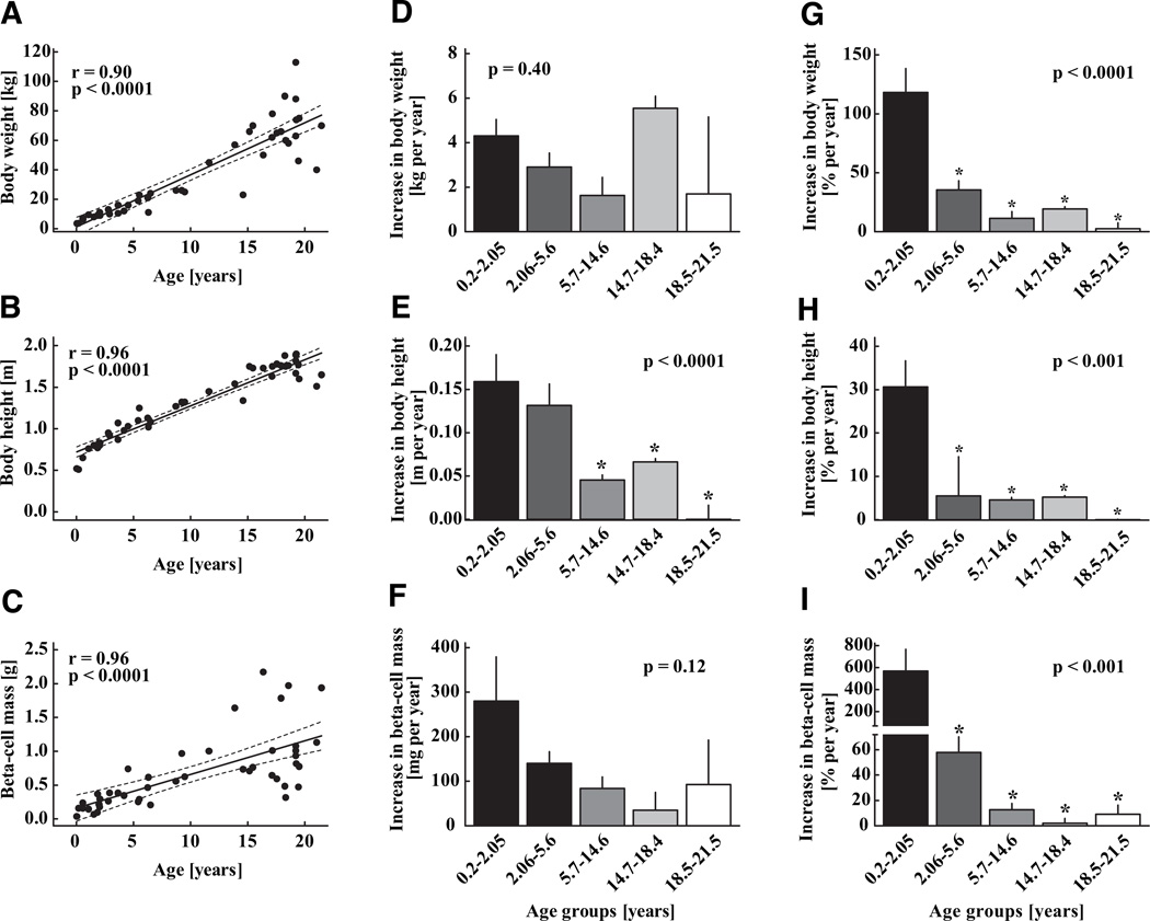
Objective: Little is known about the capacity, mechanisms, or timing of growth in beta-cell mass in humans. We sought to establish if the predominant expansion of beta-cell mass in humans occurs in early childhood and if, as in rodents, this coincides with relatively abundant beta-cell replication. We also sought to establish if there is a secondary growth in beta-cell mass coincident with the accelerated somatic growth in adolescence.

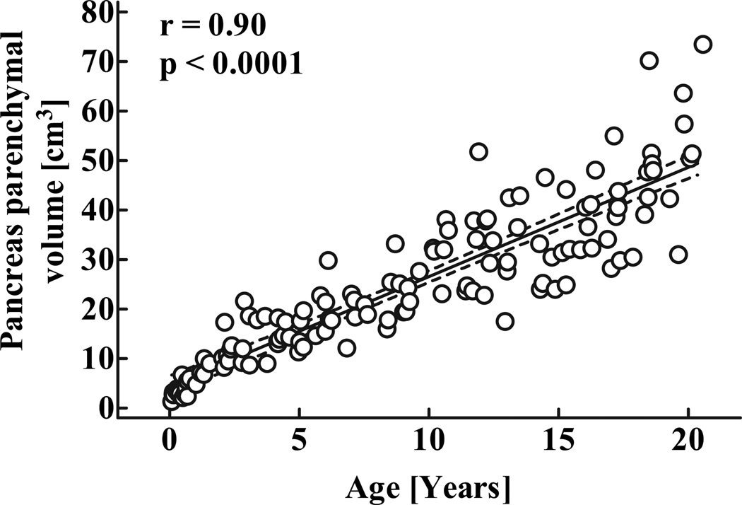
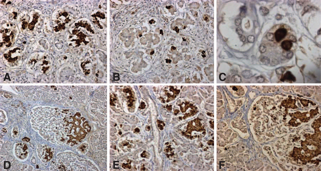
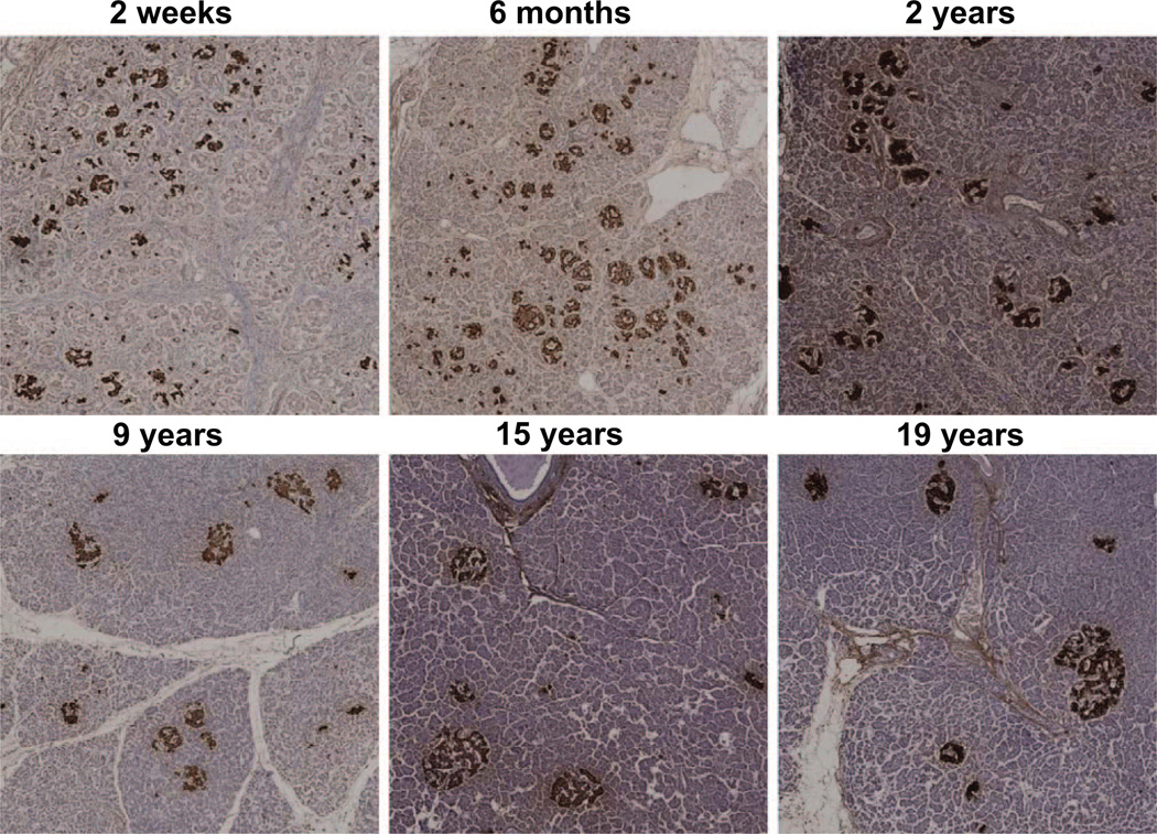
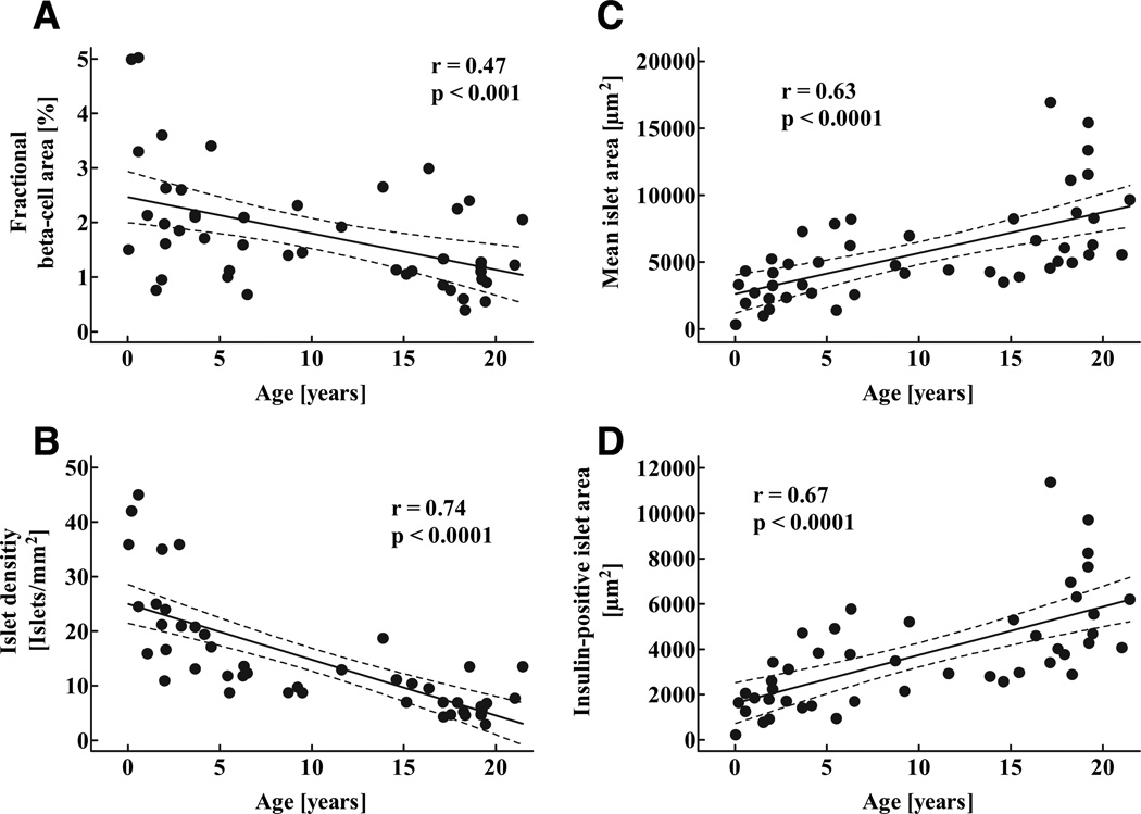
Research design and methods: To address these questions, pancreas volume was determined from abdominal computer tomographies in 135 children aged 4 weeks to 20 years, and morphometric analyses were performed in human pancreatic tissue obtained at autopsy from 46 children aged 2 weeks to 21 years.

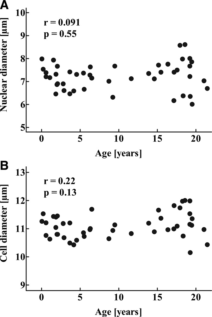
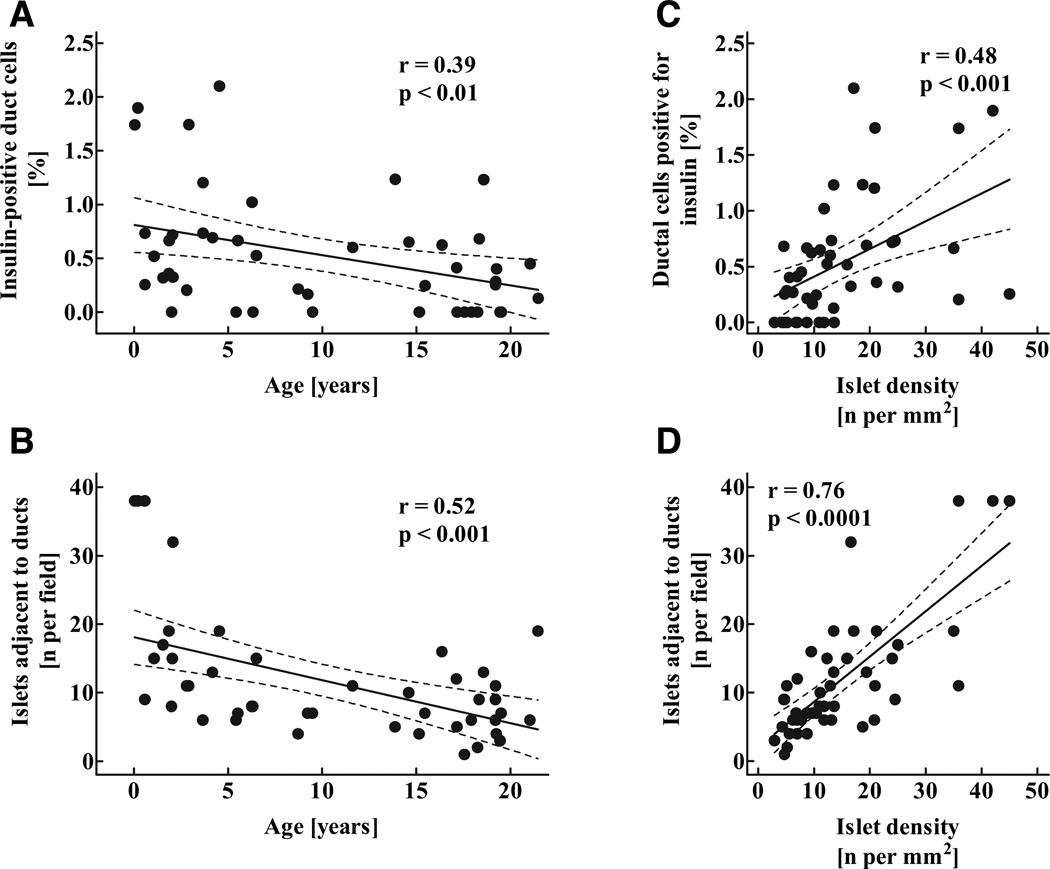
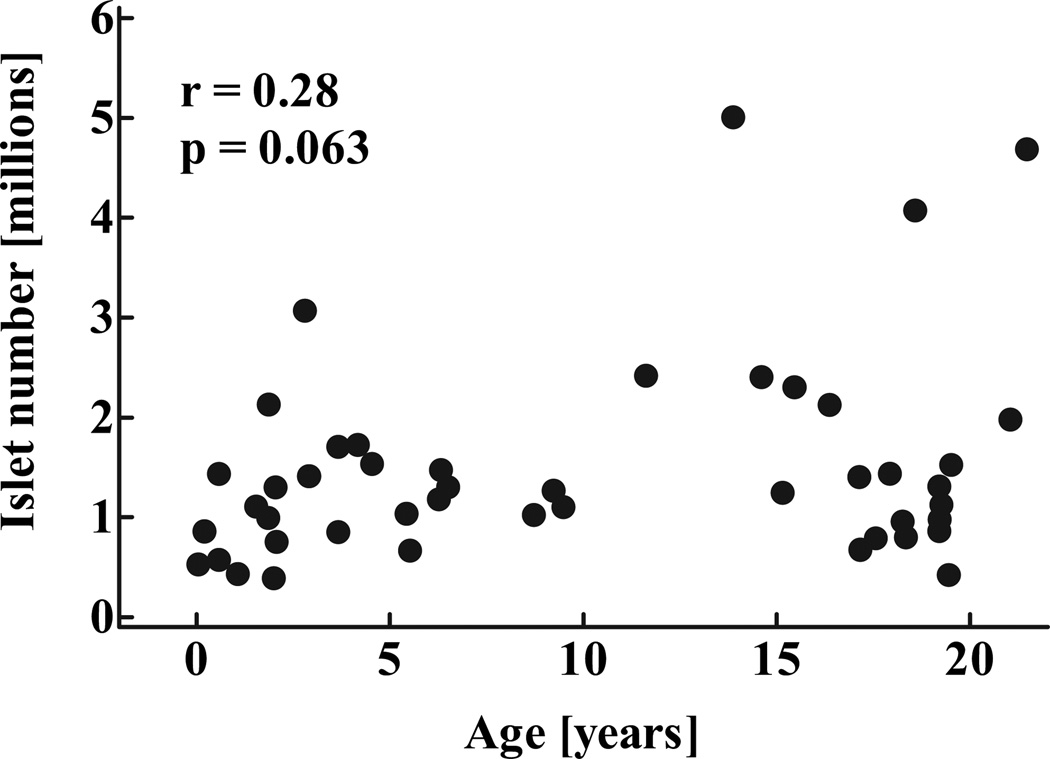
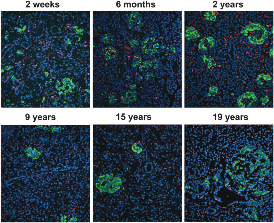
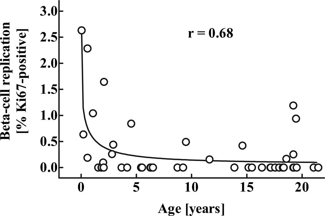
Results: We report that 1) beta-cell mass expands by severalfold from birth to adulthood, 2) islets grow in size rather than in number during this transition, 3) the relative rate of beta-cell growth is highest in infancy and gradually declines thereafter to adulthood with no secondary accelerated growth phase during adolescence, 4) beta-cell mass (and presumably growth) is highly variable between individuals, and 5) a high rate of beta-cell replication is coincident with the major postnatal expansion of beta-cell mass.

Conclusions: These data imply that regulation of beta-cell replication during infancy plays a major role in beta-cell mass in adult humans.


Figures

FIG. 1
FIG. 1
Parenchymal volume of the pancreas in 135 children aged 30 days to 20 years. Dashed lines denote the respective upper and lower 95% CIs r and P values were calculated by linear regression analysis.
FIG. 2
FIG. 2
Representative pancreatic sections stained for insulin (brown) and hematoxylin from case 1, aged 2.5 weeks (Table 1) (A–C) and case 2, aged 10 weeks (D–F), In early infancy, β-cells were abundant as small clusters (A, objective ×20) surrounded by a single cell layer nonfibrous capsule. In other areas, β-cells were mostly present as single cells (B, objective ×20), shown in high power in C (objective ×100). By 10 weeks of age, the small clusters of β-cells had grown in size and typically occupied an encapsulated islet-like structure, with the β-cells being predominantly polar within the islet as previously described (D, objective ×10; F, objective ×20). The capsule by 10 weeks had some fibrous tissue. At 10 weeks of age, islets were densely located throughout the pancreas but in higher density in some locations (E, objective ×20), in this case decorating a ductal tree.
FIG. 3
FIG. 3
Representative pancreatic sections stained for insulin (brown) and hematoxylin from six children aged 2 weeks to 19 years. Images were taken at 100 × magnification (10× objective).
FIG. 4
FIG. 4
Fractional pancreatic area positive for insulin (A), mean islet density (number of islets per mm2 pancreatic tissue (B), as well as mean islet area (C) and mean insulin-positive islet area (D) in 46 children aged 2 weeks to 21 years. Solid lines indicate the regression lines; dashed lines denote the respective upper and lower 95% CIs r and P values were calculated by linear regression analysis.
FIG. 5
FIG. 5
Nuclear diameter (A) and total diameter (B) of β-cells in 46 children aged 2 weeks to 21 years r and P values were calculated by linear regression analysis.
FIG. 6
FIG. 6
Percentage of exocrine ductal cells positive for insulin (A), mean number of islets adjacent to (five or less nuclei away) exocrine ducts (B) in 46 children aged 2 weeks to 21 years, as well as correlation between the percentage of ductal cells positive for insulin (C) and the number of islets adjacent to ducts (D) and the mean islet density. Solid lines indicate the regression lines; dashed lines denote the respective upper and lower 95% CIs r and P values were calculated by linear regression analysis.
FIG. 7
FIG. 7
Total calculated number of islets in 46 children aged 2 weeks to 21 years. Data were computed from evaluation of islet density in pancreas samples and population pancreatic volumes (Fig. 1). Data are presented as individual data points r and P values were calculated by linear regression analysis.
FIG. 8
FIG. 8
Body weight (A), body height (B), and β-cell mass (C) in 46 children aged 2 weeks to 21 years, shown as individual data. Solid lines indicate the regression lines; dashed lines denote the respective upper and lower 95% CIs r and P values were calculated by linear regression analysis. The absolute and relative increase in each parameter is shown as means ± SE on the middle and right panels (D–I) respectively. For these analyses, the group was divided into equal quintiles. Asterisks indicate significant differences versus the youngest age-group, respectively.
FIG. 9
FIG. 9
Representative pancreatic sections (same cases as in Fig. 2) stained for insulin (green), Ki67 (red), and 4′,6-diamidino-2-phenylindole dihydrochloride (DAPI) (blue) from six children aged 2 weeks to 19 years. Images were taken at 200 × magnification (20× objective).
FIG. 10
FIG. 10
Frequency of β-cell replication (percent β-cells positive for Ki67) in 46 children aged 2 weeks to 21 years. The r value was calculated using nonlinear regression analysis.

References

    1. Meier JJ, Butler PC. Insulin secretion. In: DeGroot LJ, Jameson JL, editors. Endocrinology. Philadelphia, PA: Elsevier Saunders; 2005. pp. 961–973.
    1. Goodner CJ, Koerker DJ, Weigle DS, McCulloch DK. Decreased insulinand glucagon-pulse amplitude accompanying beta-cell deficiency induced by streptozocin in baboons. Diabetes. 1989;38:925–931. - PubMed
    1. Kjems LL, Kirby BM, Welsh EM, Veldhuis JD, Straume M, McIntyre SS, Yang D, Lefebvre P, Butler PC. Decrease in beta-cell mass leads to impaired pulsatile insulin secretion, reduced postprandial hepatic insulin clearance, and relative hyperglucagonemia in the minipig. Diabetes. 2001;50:2001–2012. - PubMed
    1. Leahy JL, Bonner-Weir S, Weir GC. Abnormal glucose regulation of insulin secretion in models of reduced B-cell mass. Diabetes. 1984;33:667–673. - PubMed
    1. Gepts W. Pathologic anatomy of the pancreas in juvenile diabetes mellitus. Diabetes. 1965;114:619–633. - PubMed

Publication types