. 2008 Apr 1;22(7):854-9.
doi: 10.1101/gad.1646208. Epub 2008 Mar 11.

Tel2 mediates activation and localization of ATM/Tel1 kinase to a double-strand break

Affiliations

Tel2 mediates activation and localization of ATM/Tel1 kinase to a double-strand break

Carol M Anderson et al. Genes Dev. .

Abstract

The kinases ATM and ATR (Tel1 and Mec1 in the yeast Saccharomyces cerevisiae) control the response to DNA damage. We report that S. cerevisiae Tel2 acts at an early step of the TEL1/ATM pathway of DNA damage signaling. We show that Tel1 and Tel2 interact, and that even when Tel1 protein levels are high, this interaction is specifically required for Tel1 localization to a DNA break and its activation of downstream targets. Computational analysis revealed structural homology between Tel2 and Ddc2 (ATRIP in vertebrates), a partner of Mec1, suggesting a common structural principle used by partners of phoshoinositide 3-kinase-like kinases.


Figures

Figure 1.
Figure 1.
Tel2 is an upstream component of the TEL1 pathway of DNA damage signaling. Note that all mec1Δ strains also contain a deletion of SML1. (A) Fivefold serial dilutions of logarithmically growing cells were plated onto media containing DNA-damaging drugs. For UV treatment, cells were irradiated at 254 nm immediately after plating. (B) α-Factor-arrested cells were treated with 5 μg/mL phleomycin, and samples were collected every 10 min for Western blots. (C) Diagram of DNA damage signaling pathways in S. cerevisiae. (D) α-Factor-arrested cells were treated with 5 μg/mL phleomycin, and samples were collected every 20 min for Western blots.
Figure 2.
Figure 2.
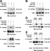
Interaction of Tel1 and Tel2. All proteins were expressed from their endogenous chromosomal loci as the only copy of the gene. (A,B) Tel1 and Tel2 constitutively interact. In each experiment, half of the doubly tagged culture was treated with 5 μg/mL phleomycin for 30 min before collection of cells. (C) Tel2-1 fails to interact with Tel1. Extracts were prepared from diploid strains TEL2/tel2-1, TEL2-Myc/tel2-1, and TEL2/tel2-1-Myc. (Top panels) Inputs and immunoprecipitations were subjected to anti-Flag immunoblotting. (Bottom panels) The same blots were stripped and reprobed with anti-Myc antibody. The faint band in the first lane is a result of Tel2-Myc protein bleeding over from the adjacent lane. (D) The Tel1–Xrs2 interaction does not depend on TEL2. More cells were used in the tel2-1 immunoprecipitations, in order to load approximately equal amounts of Flag-Tel1 protein in each immunoprecipitation. (E) The Tel1–Tel2 interaction does not depend on XRS2. Note that expression levels of Flag-Tel1 were lower in xrs2Δ strains.
Figure 3.
Figure 3.
Effects of tel2-1 on Tel1 and Tel2 levels and distribution (A) Tel1 levels are lower in tel2-1 cells. (Top panel) Four isolates of each genotype (TEL2 or tel2-1) containing Flag-Tel1 were analyzed. Equal quantities (as measured by OD600) of logarithmically growing cells were collected, and whole-cell extracts were prepared by TCA precipitation and subjected to anti-Flag immunoblotting. The same samples were run on a separate gel, and the blot was probed with anti-PGK1 antibody as a loading control. (Bottom panel) TEL2 extract was diluted with SDS-PAGE loading buffer to various final concentrations (for example, “80%” indicates the sample was diluted to 80% of its original concentration, which is a 4:5 dilution). Dilutions were run alongside undiluted tel2-1 extract and subjected to anti-Flag immunoblotting. The blot was stripped and reprobed with anti-tubulin antibody as a loading control. (B) Tel2 protein levels are not altered by the tel2-1 mutation. Four diploid TEL2-HA/tel2-1 and five diploid TEL2/tel2-1-HA strains were analyzed. Logarithmically growing cells were collected and whole-cell extracts were prepared by bead-beating in urea buffer. Equal amounts of total protein were loaded in each lane and subjected to anti-HA immunoblotting. The blot was stripped and reprobed with anti-tubulin antibody as a loading control. (C) tel2-1 does not affect the nuclear-cytoplasmic distribution of Tel1. Logarithmically growing cells were homogenized to yield whole-cell extract (W), then separated into nuclear (N) and cytoplasmic (C) fractions by differential centrifugation. Fractions were analyzed by immunoblotting with anti-Flag, anti-PGK1, or anti-nuclear pore complex (NPC) antibodies.
Figure 4.
Figure 4.
Tel1 localization to a DSB depends on its interaction with Tel2. A single DSB was induced in α-factor-arrested cells by addition of 2% galactose for 1 h. All strains contained a recognition site for HO endonuclease integrated at the TRP5 locus and a plasmid expressing HO endonuclease under the control of a galactose-inducible promoter; the endogenous HO recognition site at MATa was mutated to MATa-inc. DNA adjacent to the break site was detected by quantitative PCR. Data shown are the average of three experiments; error bars represent standard deviation of the mean. (A) Tel1 localization to a DSB is reduced in a tel2-1 strain. (B) Reduced localization of Tel1 to a DSB is not a result of lower expression levels. An extra copy of Flag-Tel1 was expressed from the CEN plasmid pRS316 in tel2-1 cells, boosting the expression level above that seen in a TEL2 strain. (Bottom panel) Western blots of cells collected just before formaldehyde fixation of cultures for ChIP. (C) Xrs2 localization to a DSB is not affected by tel2-1.

References

    1. Abraham R.T. Cell cycle checkpoint signaling through the ATM and ATR kinases. Genes & Dev. 2001;15:2177–2196. - PubMed
    1. Ahmed S., Alpi A., Hengartner M.O., Gartner A. C. elegans RAD-5/CLK-2 defines a new DNA damage checkpoint protein. Curr. Biol. 2001;11:1934–1944. - PubMed
    1. Alcasabas A.A., Osborn A.J., Bachant J., Hu F., Werler P.J., Bousset K., Furuya K., Diffley J.F., Carr A.M., Elledge S.J. Mrc1 transduces signals of DNA replication stress to activate Rad53. Nat. Cell Biol. 2001;3:958–965. - PubMed
    1. Brachmann C.B., Davies A., Cost G.J., Caputo E., Li J., Hieter P., Boeke J.D. Designer deletion strains derived from Saccharomyces cerevisiae S288C: A useful set of strains and plasmids for PCR-mediated gene disruption and other applications. Yeast. 1998;14:115–132. - PubMed
    1. Collis S.J., Barber L.J., Clark A.J., Martin J.S., Ward J.D., Boulton S.J. HCLK2 is essential for the mammalian S-phase checkpoint and impacts on Chk1 stability. Nat. Cell Biol. 2007;9:391–401. - PubMed

Publication types

MeSH terms

Substances