. 2008 Mar 18;105(11):4352-7.
doi: 10.1073/pnas.0712276105. Epub 2008 Mar 11.

p72 DEAD box RNA helicase is required for optimal function of the zinc-finger antiviral protein

Affiliations

p72 DEAD box RNA helicase is required for optimal function of the zinc-finger antiviral protein

Guifang Chen et al. Proc Natl Acad Sci U S A. .

Abstract

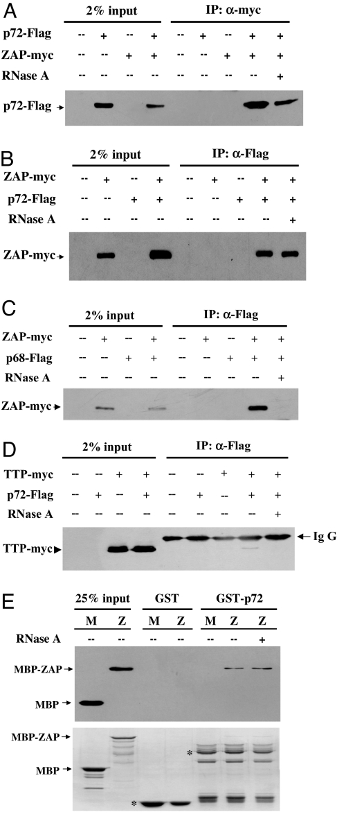
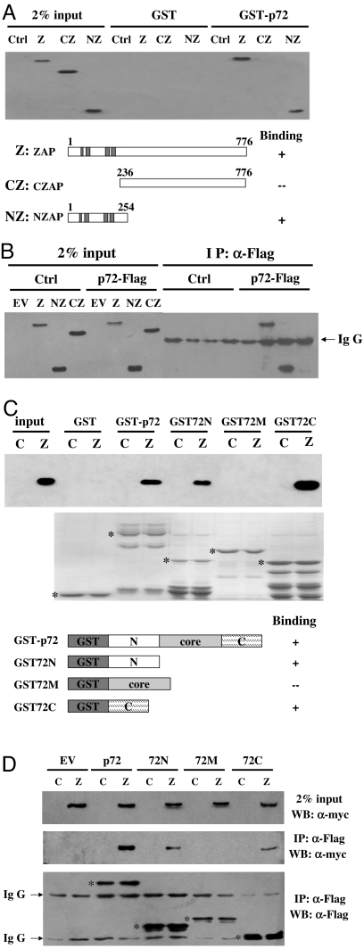
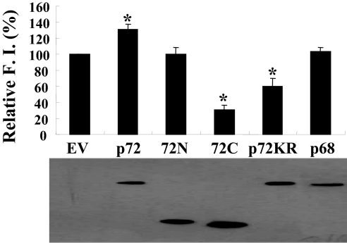
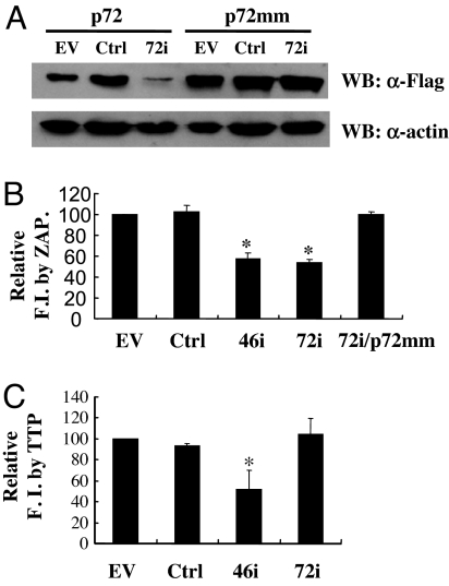
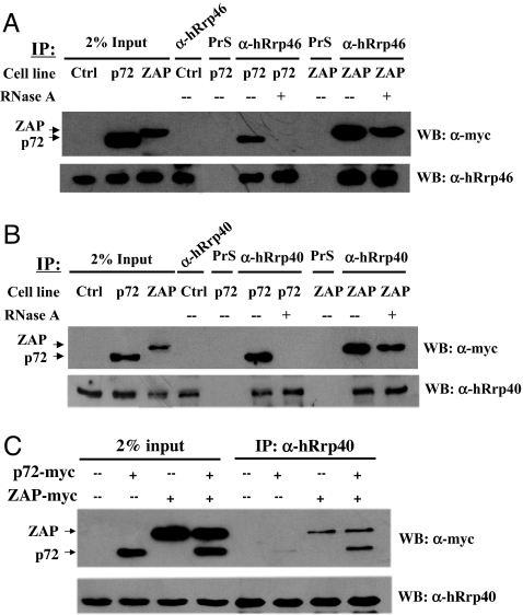
The zinc-finger antiviral protein (ZAP) specifically inhibits the replication of many viruses by preventing the accumulation of viral mRNAs in the cytoplasm. ZAP directly binds to the viral mRNAs and recruits the RNA exosome to degrade the target RNA. In the present study, we identified the p72 DEAD box RNA helicase, but not the highly similar RNA helicase p68, as a ZAP-interacting protein. The binding domain of ZAP was mapped to its N-terminal portion, whereas both the N- and C-terminal domains of p72 bound to ZAP. Overexpression of the C-terminal domain of p72 reduced ZAP's activity, whereas overexpression of the full-length p72 enhanced ZAP's activity. The RNA helicase activity was required for p72 to promote ZAP-mediated RNA degradation. Depletion of p72 by RNAi also reduced ZAP's activity but did not affect tristetraprolin-mediated RNA degradation. We conclude that p72 is required for the optimal activity of ZAP, and we propose that p72 helps to restructure the ZAP-bound target mRNA for efficient degradation.


Conflict of interest statement

The authors declare no conflict of interest.

Figures

Fig. 1.
Fig. 1.
ZAP interacts with the p72 RNA helicase. (A) The plasmids expressing myc-tagged ZAP and Flag-tagged p72 were transiently cotransfected into HEK293T cells. Forty-eight hours after transfection, the cells were lysed. The cell lysates were immunoprecipitated with the anti-myc antibody in the presence (+) or absence (−) of RNase A and Western blotted with the anti-Flag antibody. (B) The plasmids expressing Flag-tagged p72 and myc-tagged ZAP were transiently cotransfected into HEK293T cells. The cell lysates were immunoprecipitated with the anti-Flag antibody in the presence (+) or absence (−) of RNase A and Western blotted with the anti-myc antibody. (C) The plasmids expressing Flag-tagged p68 and myc-tagged ZAP were transiently cotransfected into HEK293T cells. The cell lysates were immunoprecipitated with the anti-Flag antibody in the presence (+) or absence (−) of RNase A and Western blotted with the anti-myc antibody. (D) The plasmids expressing Flag-tagged p72 and myc-tagged TTP were transiently cotransfected into HEK293T cells. The cell lysates were immunoprecipitated with the anti-Flag antibody in the presence (+) or absence (−) of RNase A and Western blotted with the anti-myc antibody. (E) Bacterially expressed GST or GST-p72 was immobilized onto glutathione-Sepharose 4B resin and incubated with bacterially expressed and partially purified myc-tagged MBP or ZAP, which was fused to myc-tag at the C terminus and to MBP at the N terminus (MBP-ZAP). The resins were washed and boiled in the sample loading buffer. The proteins were resolved by SDS/PAGE and detected by Western blotting with the anti-myc antibody (Upper) or by Coomassie blue staining (Lower). The positions of MBP and MBP-ZAP are indicated by arrows, and the positions of GST and GST-p72 are indicated by asterisks. M, MBP; Z, MBP-ZAP.
Fig. 2.
Fig. 2.
Mapping the binding domains of ZAP and the p72 RNA helicase. (A) Bacterially expressed GST or GST-p72 was immobilized onto glutathione-Sepharose 4B resin and incubated with the lysates of the cells expressing the indicated myc-tagged ZAP proteins in the presence of RNase A at 4°C for 2 h. The resins were washed and boiled in the sample loading buffer. (Upper) The proteins were resolved by SDS/PAGE and detected by Western blotting using the anti-myc antibody. (Lower) Schematic representations of the ZAP proteins are shown, and their binding activities are summarized. Input, total cell lysate; Ctrl, control cells. (B) The plasmid expressing the indicated myc-tagged ZAP protein and the plasmid expressing Flag-tagged p72 were transiently cotransfected into HEK293T cells. The cell lysates were immunoprecipitated with the anti-Flag antibody in the presence of RNase A and Western-blotted with the anti-myc antibody. Input, total cell lysate; Ctrl, control plasmid pCMVHF; EV, empty vector pcDNA4. (C) Bacterially expressed recombinant proteins of the indicated p72-truncation mutants were immobilized onto glutathioneSepharose 4B resin and incubated with the lysates of control cells or ZAP-expressing cells in the presence of RNase A at 4°C for 2 h. The resins were washed and boiled in the sample loading buffer. (Upper) The proteins were resolved by SDS/PAGE and detected by Western blotting using the anti-myc antibody. (Lower) Schematic representations of the p72 proteins are shown and their binding activities are summarized. Input, total cell lysate; C, control cells; Z, ZAP-expressing cells. The positions of the GST proteins are indicated by asterisks. (D) The plasmid expressing the indicated Flag-tagged p72 proteins were transiently transfected into ZAP-expressing cells or control cells. The cell lysates were immunoprecipitated with the anti-Flag antibody in the presence of RNase A and Western-blotted with the indicated antibodies. Input, total cell lysate; C, control cells; Z, ZAP-expressing cells. EV, empty vector. The positions of the p72 proteins are indicated by asterisks.
Fig. 3.
Fig. 3.
Effects of the overexpression of the p72 mutants on ZAP's activity. The plasmid expressing the indicated Flag-tagged p72 proteins or Flag-tagged p68 was cotransfected with the pMLV-luc reporter into 293TRex-ZAP cells. Immediately after transfection, tetracycline was added to induce ZAP expression. Forty-eight hours after transfection, the cells were lysed. An aliquot of the lysate was analyzed for the luciferase activities, and another aliquot was analyzed for the expression of the p72 proteins or p68 by Western blotting. Fold inhibition was calculated as the luciferase activity in the mock-treated cells divided by the luciferase activity in the tetracycline-treated cells. (Upper) Relative fold inhibition was calculated as the fold inhibition in the presence of empty vector divided by the fold inhibition in the presence of the p68 or p72 proteins. The relative fold inhibition data are means + SE of three independent experiments, and the Western blotting result (Lower) is representative of three independent experiments. The asterisks denote to P < 0.05. F.I., fold inhibition; EV, empty vector; 72N, N-terminal domain of p72; 72C, C-terminal domain of p72; p72KR, p72-K142R mutant.
Fig. 4.
Fig. 4.
Depletion of p72 by RNAi reduced ZAP's activity. (A) The plasmid expressing Flag-tagged p72 or the p72 rescue plasmid (p72 mm) was cotransfected into 293TRex cells with the plasmid expressing control shRNA (Ctrl) or the shRNA directed against p72 (72i). The expression levels of the p72 proteins were judged by Western blotting using anti-Flag and anti-β-actin antibodies. (B) The pMLV-luc reporter was cotransfected with the plasmid expressing the indicated shRNA or 72i plus p72 mm into 293TRex-ZAP cells. Immediately after transfection, tetracycline was added to induce ZAP expression. Forty-eight hours after transfection, the luciferase activities were measured. Fold inhibition was calculated as in Fig. 3. Relative fold inhibition was calculated as the fold inhibition in the absence of the shRNA (empty vector) divided by the fold inhibition in the presence of the indicated RNAi. The data are means + SE of four independent experiments. The asterisks denote P < 0.05. (C) The ARE-containing reporter, pGL3-ARE-Luc, was cotransfected with the plasmid expressing the indicated shRNA into 293TRex-TTP cells. Immediately after transfection, tetracycline was added to induce TTP expression. Forty-eight hours after transfection, the luciferase activities were measured. Fold inhibition was calculated as the luciferase activity in the mock-treated cells divided by the luciferase activity in the tetracycline-treated cells. Relative fold inhibition was calculated as the fold inhibition in the absence of the shRNA (empty vector) divided by the fold inhibition in the presence of the indicated shRNA. The data are means + SE of three independent experiments. The asterisks denote to P < 0.05. EV, empty vector; 46i, shRNA-directed against the exosome component hRrp46; FI, fold inhibition.
Fig. 5.
Fig. 5.
The p72 RNA helicase does not directly interact with the exosome. (A and B) The 293TRex cells expressing myc-tagged p72 or myc-tagged ZAP were treated with tetracycline to induce ZAP or p72 expression and lysed in the lysis buffer in the presence (+) or absence (−) of 50 μg/ml RNase A. The proteins were immunoprecipitated with anti-hRrp46p (A) or anti-hRrp40p (B) antibody and Western blotted with the indicated antibodies. Input, total cell lysate; Ctrl, 293TRex cells; p72, 293TRex cells expressing p72; ZAP, 293TRex cells expressing ZAP. (C) The HEK293T cells expressing myc-tagged p72, myc-tagged ZAP, or both were lysed in the lysis buffer in the presence of 50 μg/ml RNase A. The proteins were immunoprecipitated with anti-hRrp40p antibody and Western blotted with the anti-myc antibody (Upper) or with the anti-hRrp40p antibody (Lower).

References

    1. Gao G, Guo X, Goff SP. Inhibition of retroviral RNA production by ZAP, a CCCH-type zinc finger protein. Science. 2002;297:1703–1706. - PubMed
    1. Müller S, et al. Inhibition of filovirus replication by the zinc finger antiviral protein. J Virol. 2006;10:1601–1606. - PMC - PubMed
    1. Bick MJ, et al. Expression of the zinc-finger antiviral protein inhibits alphavirus replication. J Virol. 2003;77:11555–11562. - PMC - PubMed
    1. Ryman KD, et al. Sindbis virus translation is inhibited by a PKR/RNase L-independent effector induced by alpha/beta interferon priming of dendritic cells. J Virol. 2005;79:1487–1499. - PMC - PubMed
    1. Guo X, et al. The zinc finger antiviral protein directly binds to specific viral mRNAs through the CCCH zinc finger motifs. J Virol. 2004;78:12781–12787. - PMC - PubMed

Publication types

Substances