. 2008 Feb;13(1):1-16.
doi: 10.3233/jad-2008-13101.

C-terminal cleavage of the amyloid-beta protein precursor at Asp664: a switch associated with Alzheimer's disease

Affiliations

C-terminal cleavage of the amyloid-beta protein precursor at Asp664: a switch associated with Alzheimer's disease

Surita Banwait et al. J Alzheimers Dis. 2008 Feb.

Abstract

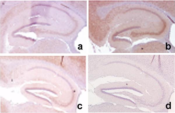
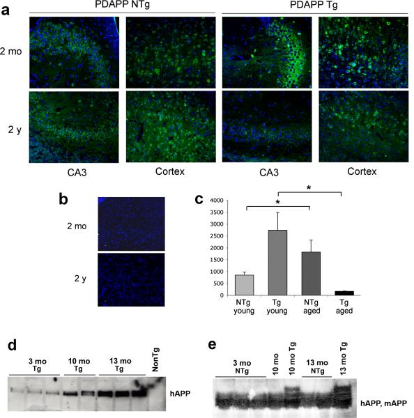
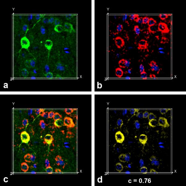
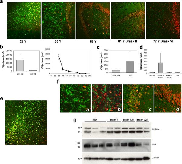
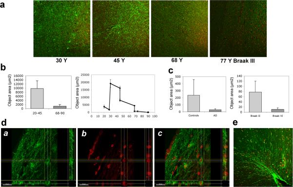
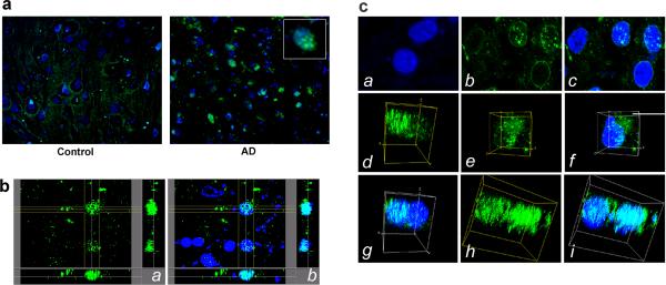
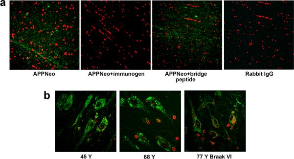
In addition to the proteolytic cleavages that give rise to amyloid-beta (Abeta), the amyloid-beta protein precursor (AbetaPP) is cleaved at Asp664 intracytoplasmically. This cleavage releases a cytotoxic peptide, APP-C31, removes AbetaPP-interaction motifs required for signaling and internalization, and is required for the generation of AD-like deficits in a mouse model of the disease. Although we and others had previously shown that Asp664 cleavage of AbetaPP is increased in AD brains, the distribution of the Asp664-cleaved forms of AbetaPP in non-diseased and AD brains at different ages had not been determined. Confirming previous reports, we found that Asp664-cleaved forms of AbetaPP were increased in neuronal cytoplasm and nuclei in early-stage AD brains but were absent in age-matched, non-diseased control brains and in late-stage AD brains. Remarkably, however, Asp664-cleaved AbetaPP was prominent in neuronal somata and in processes in entorhinal cortex and hippocampus of non-diseased human brains at ages <45 years. Our observations suggest that Asp664 cleavage of AbetaPP may be part of the normal proteolytic processing of AbetaPP in young (<45 years) human brain and that this cleavage is down-regulated with normal aging, but is aberrantly increased and altered in location in early AD.


Figures

Fig. 1
Fig. 1
Cleavage of hAPP at Asp664 in vivo is abolished in PDAPP(D664A) transgenic mice. A rabbit antibody specific for the neoepitope generated after cleavage of AβPP at Asp664, APPNeo [,,–17,63], was used to stain hippocampal sections of a, non-transgenic; b, PDAPP transgenic and c, PDAPP(D664A) transgenic mice. d, APPNeo antiserum was replaced by preimmune sera.
Fig. 2
Fig. 2
Cleavage of transgenic hAPP and endogenous mAPP are differentially regulated by aging. a, Representative confocal images of young [2 months of age (mo)] and aged [2 years of age (2 y)] mouse brain sections immunostained with APPNeo b, APPNeo was replaced by preimmune rabbit sera. Sections were counterstained with TOTO-3 to visualize nuclei. The far red channel was pseudocolored blue. c, Total area of APPNeo immunoreactivity was quantified in hippocampal sections as described in Methods. d, Lysates from mouse brains as indicated were probed with an antibody that recognizes the extracellular domain of hAPP (5A3/1G7). e, Lysates from mouse brains as indicated were probed with an antibody that recognizes both hAPP and mAPP (CT15).
Fig. 3
Fig. 3
Asp664-cleaved hAPP are associated with the ER. a, APPNeo (green channel) and α-PDI (red channel) antibodies were used in double immunofluorescence assays on transgenic mouse brain sections followed by Alexa488- and Alexa555-conjugated α-rabbit and α-mouse secondary antibodies respectively. TOTO-3 was used as counterstain to visualize nuclei. The far red channel was pseudocolored blue. Snapshots from maximum intensity projection Imaris Surpass volume images of a representative confocal z-stack for each channel (a, APPNeo, green; b, PDI, red; c, overlap; d, colocalization channel) are shown.
Fig. 4
Fig. 4
Asp664 cleavage in human hippocampus. a, Representative low-magnification (100X) confocal images of hippocampal sections from patients at the indicated ages, stained with APPNeo and counterstained with TOTO-3 to visualize nuclei. Braak stages are denoted for AD cases. b and c, Total areas of APPNeo immunoreactivity in non-diseased and AD human samples were quantified as described in Methods. b, Left panel, averages ± SEM of young (20−45 Y) and aged (68−90 Y) are shown. Right panel, averages ± SEM of the indicated ages are shown. c, Total area of APPNeo immunoreactivity in control and AD groups. d, Total area of APPNeo immunoreactivity in different Braak stage groups within the AD group were compared to controls. Averages ± SEM are shown. AD, no break stage specified. e, Representative low-magnification confocal image of APPNeo-immunostained hippocampus in a 23 Y patient. TOTO-3 was used to counterstain nuclei. f, Representative high-magnification confocal images of CA3 hilus (a and b) and ML granular layer (c and d) in young (a and c) and aged (b and d) non-diseased human brains. g, Lysates from tissue samples obtained from the indicated groups were separated electrophoretically and reacted with the indicated antibodies. ND, non diseased.
Fig. 5
Fig. 5
Asp664 cleavage in human entorhinal cortex. a, Representative low-magnification (100X) confocal images of hippocampal sections from patients at the indicated ages, stained with APPNeo and counterstained with TOTO-3 to visualize nuclei. Braak stage is denoted for the AD case. b and c, Total areas of APPNeo immunoreactivity in non-diseased and AD human samples were quantified as described in Methods. b, Left panel, averages ± SEM of young (20−45 Y) and aged (68−90 Y) samples are shown. Right panel, averages ± SEM of the indicated ages are shown. c, Left panel, total area of APPNeo immunoreactivity in control and AD groups. Right panel, total area of APPNeo immunoreactivity in different Braak stage groups within the AD group were compared to controls. Averages ± SEM are shown. d, Section views of a, green; b, red; c, overlay channels in stacks of confocal images collected from dorsal EC in brain sections of a 30 Y old non-diseased patient immunostained with APPNeo. f, Representative low-magnification (100X) confocal image of parahippocampal gyrus in brain sections of a 45 Y non-diseased patient immunostained with APPNeo. Sections were counterstained with TOTO-3 to visualize nucleicaption.
Fig. 6
Fig. 6
Asp664-cleaved AβPP fragments form punctate foci in nuclei of hippocampal neurons in AD brain. a, representative high-magnification (600X) confocal images of hippocampal brain sections from control and AD brains stained with APPNeo and counterstained with TOTO-3. Inset, digital magnification of a representative nucleus containing APPNeo-immunoreactive material. b, Section views of stacks of confocal images from AD brain stained with APPNeo (a) and counterstained with TOTO-3 (b). The regions of colocalization of the green and blue channels in (b) are shown in cyan. c, Representative high-magnification (2000X: 1000X magnification × 2X digital zoom) confocal images (a, b, c) or snapshots of maximum intensity projections Surpass volume images (d through i) of green and blue channels in stacks of confocal images of representative nuclei in non-diseased (a) or AD brain sections (b through i). APPNeo-immunoreactive material decorates the perinuclear region and discrete nuclear foci (b and c), in a region juxtaposed to the nucleus (e and f), or is associated with large regions of chromatin (d and g, h and i) in hippocampal neurons of AD brain.
Fig. 7
Fig. 7
APPNeo specificity and integrity of human tissue. a, APPNeo staining is abolished if the APPNeo antiserum is preadsorbed with the immunogen peptide or if it is replaced by preimmune rabbit IgG. Immunostaining is unaffected when the APPNeo antiserum is preadsorbed with a peptide encompassing the Asp664 cleavage site and the first four amino acids of AβPP (bridge peptide, Methods). b, Calreticulin immunostaining reveals that the overall cellular structure and tissue integrity are preserved in AD samples. TOTO-3 was used to counterstain nuclei.

Similar articles

Cited by

References

    1. Artavanis-Tsakonas S, Matsuno K, Fortini ME. Notch signaling. Science. 1995;268:225–232. - PubMed
    1. Ayala-Grosso C, Ng G, Roy S, Robertson GS. Caspase-cleaved amyloid precursor protein in Alzheimer's disease. Brain Pathol. 2002;12:430–441. - PMC - PubMed
    1. Baek SH, Ohgi KA, Rose DW, Koo EH, Glass CK, Rosenfeld MG. Exchange of N-CoR corepressor and Tip60 coactivator complexes links gene expression by NF-kappaB and beta-amyloid precursor protein. Cell. 2002;110:55–67. - PubMed
    1. Besnault-Mascard L, Leprince C, Auffredou MT, Meunier B, Bourgeade MF, Camonis J, Lorenzo HK, Vazquez A. Caspase-8 sumoylation is associated with nuclear localization. Oncogene. 2005;24:3268–3273. - PubMed
    1. Biederer T, Cao X, Sudhof TC, Liu X. Regulation of APP-dependent transcription complexes by Mint/X11s: differential functions of Mint isoforms. J Neurosci. 2002;22:7340–7351. - PMC - PubMed

Publication types

MeSH terms