. 2008 May;332(2):227-35.
doi: 10.1007/s00441-008-0588-6. Epub 2008 Mar 12.

Early markers of regeneration following ductal ligation in rat submandibular gland

Affiliations

Early markers of regeneration following ductal ligation in rat submandibular gland

Emanuele Cotroneo et al. Cell Tissue Res. 2008 May.

Abstract

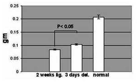
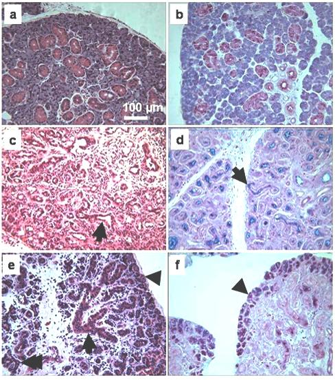
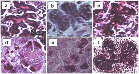
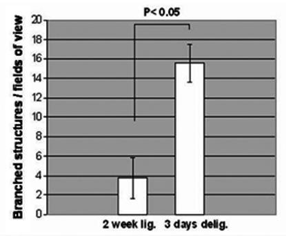
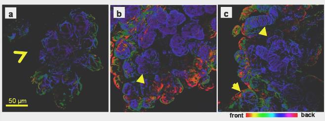
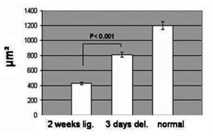
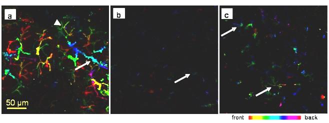
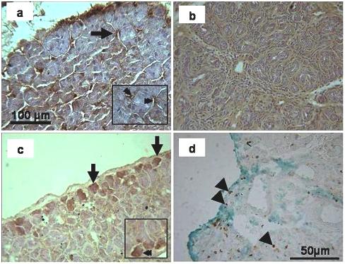
Rat submandibular glands can recover their function and secretory protein content following ductal ligation-induced atrophy. Morphological studies have established that following ligation, deligation of the gland allows the regeneration of new salivary gland tissue. However, little is known about changes happening during early regeneration following intra-oral duct ligation, which does not damage the parasympathetic nerves. Glands that had been 2 weeks ligated or 2 weeks ligated + 3 days deligated were compared. Tissue was prepared for histological, immunohistochemical (SMG-B and Ki-67) and immunocytochemical analyses (smooth muscle actin, aquaporin 5). Haematoxylin and eosin staining of deligated glands showed that some acini regained their cytoplasmic volume; moreover, the loss of Alcian blue/periodic acid-Schiff's staining from the lumen of ducts suggested successful deligation. The deligated gland was characterized by atypical acinar-ductal branched structures, which were less frequent in the ligated gland and rarely seen in normal unoperated tissue. Myoepithelial cells were also investigated since changes in their morphology reflected changes in the acini morphology not readily detected by conventional staining. Actin staining revealed the presence of some shrunken acini in the atrophic tissue, whereas they had regained their normal morphology in the deligated gland suggesting that the acini were recovering. Some acini during deligation regained aquaporin 5 expression, which had decreased during atrophy. SMG-B protein, located in the pro-acinar cell during gland development and usually found in the intercalated duct cells in the adult, was detected in the newly formed acini of the deligated gland. This study suggests that morphological markers of regeneration appear as early as 3 days following ligation removal.


Figures

Fig. 1
Fig. 1
Mean weight (± SEM) of 2 weeks ligated (2 weeks lig.), of 2 weeks ligated plus 3 days de-ligated (3 days del.) and unoperated (normal) adult submandibular glands. The 2 weeks ligated plus 3 days de-ligated gland (n=5) showed an increase in weight (p < 0.05) compared to the 2 weeks ligated (n=5), but were nevertheless half the typical weight of a normal gland (n=4).
Fig. 2
Fig. 2
Histological comparison among unoperated gland, ligated, and de-ligated submandibular gland. a, b) Unoperated gland H&E and AB/PAS, respectively, showing typical appearance of acinar and duct cells. c) H&E stained section of ligated gland, showing luminal dilatation of the duct (arrow), absence of acini and extensive inflammation. d) AB/PAS in ligated gland, showing loss of cellular secretory granules and material in the lumen of the ducts (arrow). e) 3 days de-ligated gland H&E section; some acini (arrow head) and ductal cells (arrow) have recovered some of their size. Acinar-ductal branched structures are often visible (double arrow). f) AB/PAS in de-ligated gland, showing some acini that have recovered their glycoproteins content (arrowhead).
Fig. 3
Fig. 3
Branched structures occurring in the 3 days deligated tissue (a, b, d, e) and in the 2 weeks ligated tissue (c). The AB/PAS staining shows presence of glycoproteins inside the immature acini at the end of the branched structures (d, e). (f) Branched structures occurring during the embryonic development of rat submandibular gland, (embryonic day 18).
Fig. 4
Fig. 4
Mean (± SEM) number of branched structures per field of view (200 × magnification) in the 2 weeks ligated (2 weeks lig.) and in the 2 weeks ligated plus 3 days de-ligated (3 days del.) submandibular gland stained by H&E. The 2 weeks plus 3 days de-ligated gland showed an increase in the frequency of the branched structures (p< 0.05, 5 observations from 5 rats, n=25) compared to the 2 weeks ligated. In the unoperated tissue, no branched structures were identified.
Fig. 5
Fig. 5
Actin immunofluorescence in adult rat submandibular gland. Collagenase-digested cells were incubated with an anti-smooth muscle actin antibody and viewed by confocal microscopy. Optical sections (approximately 1μm) were taken and projected to create a 3D image and colour-coded according to the depth of field. a) Unoperated submandibular gland, star-shaped myoepithelial cells surround the acini, but not the big ducts (open arrowhead). b) In the ligated gland myoepithelial cells have lost their typical morphology but reveal the presence of numerous shrunken acini (arrowhead). c) In the de-ligated gland the myoepithelial cells on the acini recovered their normal star-shaped morphology (arrow). In ligated and deligated tissue myoepithelial cells were found on the big ducts (arrow head).
Fig. 6
Fig. 6
Mean (± SEM) area of the acini from the 2 weeks ligated (2 weeks lig.), from the 2 weeks ligated plus 3 days de-ligated (3 days del.) and from unoperated (normal) submandibular gland. The 3 days de-ligated gland showed a significant increase in the acini area (p < 0.001, 20 observations from 3 samples n=60) compared to the 2 weeks ligated only glands.
Fig. 7
Fig. 7
AQP5 immunofluorescence in rat submandibular gland. Collagenase-digested cells were incubated with an anti-AQP5. All images were taken using the same settings; different colours represent differences in depth of field. a) In the control gland AQP5 is strongly expressed along the apical membrane, the intercellular secretory canaliculi (arrowhead) and along the intercalated ducts (arrow). b) In the ligated tissue weak AQP5 expression is restricted to the apical membrane of presumed acinar cells (arrow). c) Some acini in the deligated tissue have regained some AQP5 expression along the intercellular secretory canaliculi (arrow)
Fig. 8
Fig. 8
SMG-B immunohistochemistry of the submandibular gland. a) In the unoperated control the staining appeared in some intercalated ducts (arrow; see also double arrow in inset) but not in the acini. b) The 2 weeks ligated gland did not show any localised staining in the parenchyma. c) In the de-ligated tissue, some acini on the edge of lobule showed positive immunoreactivity (arrow; see also double arrow in inset). d) Ki-67 staining (counterstained with Light Green) on 3 day de-ligated plus 2 week ligated submandibular gland. Some cells on the edge of lobules (arrowheads) and in the centre of glands (arrowheads) were labelled.

References

    1. Ball WD, Hand AR, Johnson AO. Secretory proteins as markers for cellular phenotypes in rat salivary glands. Dev Biol. 1988;125:265–279. - PubMed
    1. Ball WD, Hand AR, Moreira JE. A neonatal secretory protein associated with secretion granule membranes in developing rat salivary glands. J Histochem Cytochem. 1991;39:1693–1706. - PubMed
    1. Borghese E. The development in vitro of the submandibular and sublingual glands of Mus musculus. J Anat. 1950;84:287–302. - PMC - PubMed
    1. Burgess K, Dardick I, Cummins MM, Burford-Mason AP, Basset R, Brown DH. Myoepithelial cells actively proliferate during atrophy of rat parotid gland. Radiol Endod. 1986;82:674–680. - PubMed
    1. Carpenter G, Osailan SM, Correia PN, Paterson KL, Proctor GB. Rat salivary gland ligation causes reversible secretory hypofunction. Acta Physiol. 2007;189:241–249. - PMC - PubMed

Publication types

LinkOut - more resources