. 2008 Mar 11;6(3):e51.
doi: 10.1371/journal.pbio.0060051.

Creatine kinase-mediated ATP supply fuels actin-based events in phagocytosis

Affiliations

Creatine kinase-mediated ATP supply fuels actin-based events in phagocytosis

Jan W P Kuiper et al. PLoS Biol. .

Abstract

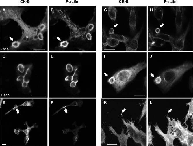
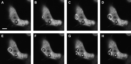
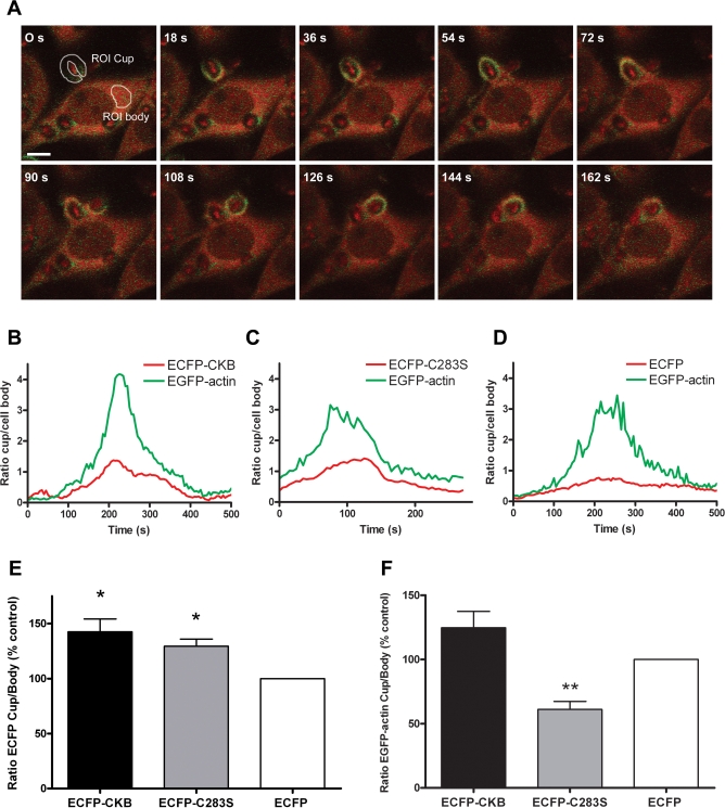
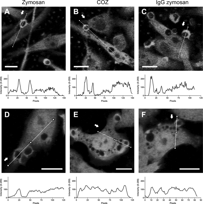
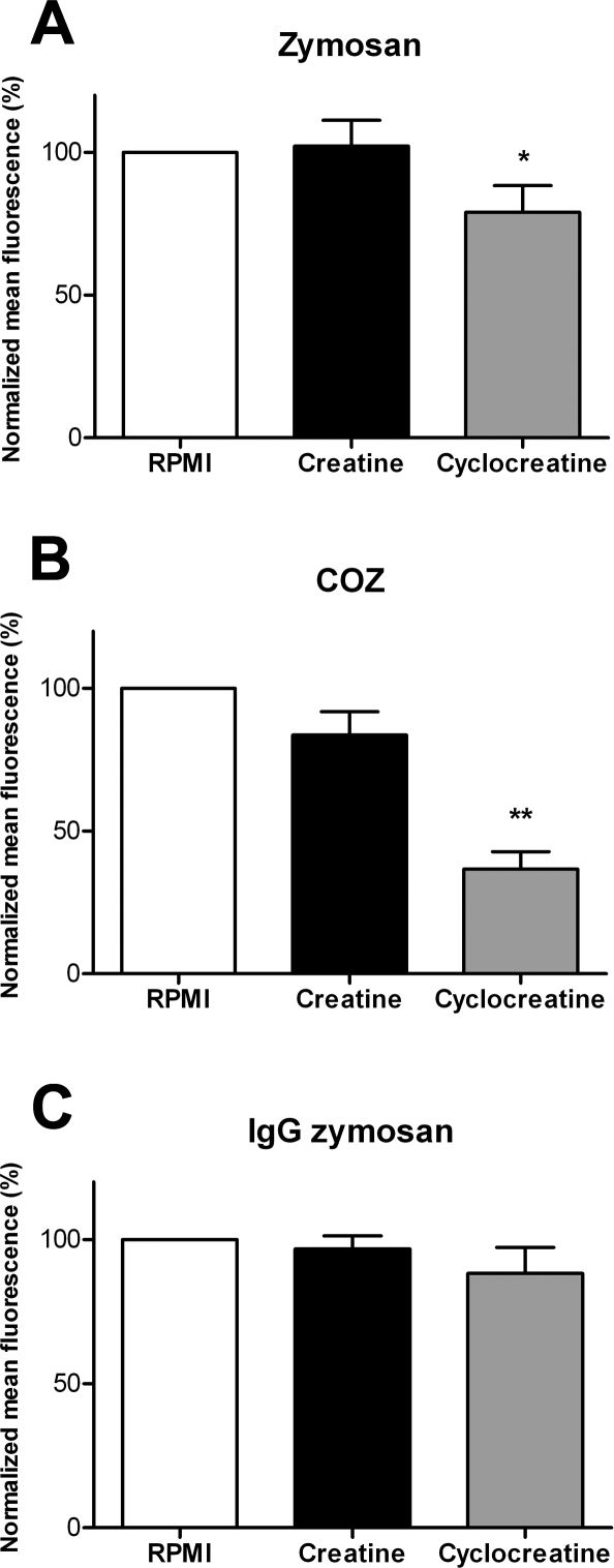
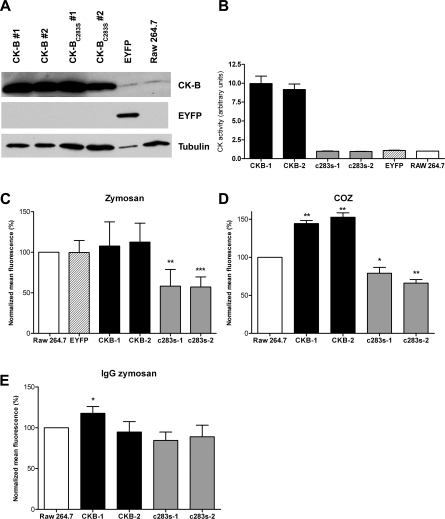
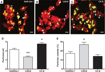
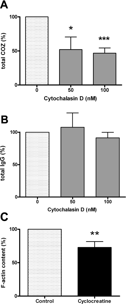
Phagocytosis requires locally coordinated cytoskeletal rearrangements driven by actin polymerization and myosin motor activity. How this actomyosin dynamics is dependent upon systems that provide access to ATP at phagosome microdomains has not been determined. We analyzed the role of brain-type creatine kinase (CK-B), an enzyme involved in high-energy phosphoryl transfer. We demonstrate that endogenous CK-B in macrophages is mobilized from the cytosolic pool and coaccumulates with F-actin at nascent phagosomes. Live cell imaging with XFP-tagged CK-B and beta-actin revealed the transient and specific nature of this partitioning process. Overexpression of a catalytic dead CK-B or CK-specific cyclocreatine inhibition caused a significant reduction of actin accumulation in the phagocytic cup area, and reduced complement receptor-mediated, but not Fc-gammaR-mediated, ingestion capacity of macrophages. Finally, we found that inhibition of CK-B affected phagocytosis already at the stage of particle adhesion, most likely via effects on actin polymerization behavior. We propose that CK-B activity in macrophages contributes to complement-induced F-actin assembly events in early phagocytosis by providing local ATP supply.


Conflict of interest statement

Competing interests. The authors have declared that no competing interests exist.

Figures

Figure 1
Figure 1. Cytosolic CK-B Accumulates in the Phagocytic Cup Area of Macrophages
Uptake of zymosan in primary microglia (A–D), primary peritoneal macrophages (E and F), and RAW 264.7 macrophages (G–J). Fixation followed by (immuno)staining with CK-B antibodies (A, C, E, G, I, and K) or phalloidin (B, D, F, H, J, and L) reveals the co-accumulation of CK-B and F-actin at the phagocytic cup (arrows). (C and D) Saponin (sap) extraction of phagocytosing microglia prior to fixation and CK-B or actin staining (I–L) RAW 264.7 macrophages overexpressing mouse CK-B. Note that CK-B shows additional pronounced spot-like accumulation at the distal tips of filopodia (arrows, [K and L]). Bar represents 10 μm.
Figure 2
Figure 2. Transient Recruitment of EYFP-Tagged CK-B
Time-lapse microscopy of zymosan uptake in RAW 264.7 cells transiently transfected with EYFP-tagged CK-B. Eight subsequent images captured at 6-s intervals over a 48-s recording period are shown. Asterisks indicate zymosan particles binding at the cellular membrane, during the process of engulfment, and after internalization. Bar indicates 5 μm.
Figure 3
Figure 3. CK-BC283S Decreases Actin Accumulation in Phagocytic Cups
(A) Time-lapse images of a cell coexpressing EGFP-actin and ECFP-CK-B in the process of internalizing a zymosan particle, demonstrating the simultaneous accumulating signals from ECFP-CK-B (red) and EGFP-actin (green). Representative regions of interest (ROIs) are being shown for the phagocytic cup (ROI cup) and the cytosol (ROI body). Bar indicates 5 μm. (B–D) Signal intensities in the phagocytic cup and cell body were analyzed in cells coexpressing EGFP-actin and ECFP-CK-B, ECFP-CK-B(C283S), or ECFP. The average pixel intensities in the ROIs were determined and the cup/body ratios for EGFP-actin (green) and ECFP-CK-B, ECFP-CK-B(C283S), or ECFP (red) plotted against time. (E) Average maximal cup/body ratios for 10–16 events in cells expressing ECFP-CK-B, ECFP-CK-B(C283S), and ECFP. (F) Average maximal EGFP-actin cup/body ratios in the same cells as in (E). Bars depict mean value with error bars representing the standard error of the mean (SEM). Number of events analyzed: n = 16 for ECFP-CK-B; n = 10 for ECFP-CK-B(C283S) and n = 13 for ECFP. *p < 0.01; **p < 0.005.
Figure 4
Figure 4. Dynamic Redistribution of EYFP-Tagged CK-B during Phagocytosis.
Time-lapse microscopy of zymosan (A and D), COZ (B and E), or IgG-opsonized zymosan (C and F) uptake in RAW 264.7 cells stably transfected with EYFP-CK-B (A–C) or EYFP (D–F). Photos represent a single frame at the peak of accumulation from a time-lapse recording; arrows mark the start of the corresponding line plot visualizing accumulation of signal in the cup. Bars indicate 10 μm.
Figure 5
Figure 5. Cyclocreatine Inhibits Phagocytosis
Fluorescent particle uptake capacity quantified by FACS in RAW 264.7 cells preincubated with 5 mM creatine, 5 mM cyclocreatine, or normal growth medium. (A) Zymosan. (B) COZ. (C) IgG-opsonized zymosan. Bars represent averages of three to four experiments performed in duplicate (±standard deviation [SD]). *p < 0.03; **p < 0.005.
Figure 6
Figure 6. CK-B and CK-B(C283S) Influence Phagocytosis
(A) Western blot analysis of RAW 264.7 cells stably expressing CK-B or CK-B(C283S). Two individual retrovirally transduced populations were maintained (CK-B#1 and CK-B#2, and CK-B(C283S)#1 and CK-B(C283S)#2). RAW 264.7 cells expressing EYFP and noninfected cells were used as control. (B) CK enzymatic activity of depicted cell lysates. Fluorescent particle uptake capacity quantified by FACS in cell lines incubated with zymosan (C), COZ (D), or IgG zymosan (E). Bars represent the average of three to four experiments (±SD). *p < 0.05; **p < 0.005; ***p < 0.0005.
Figure 7
Figure 7. CK-B Is Involved in the Initial Steps of Phagocytosis
Adhesion and uptake of FITC-labeled COZ particles in RAW 264.7 cells (A) and RAW 264.7 cells expressing CK-B(C283S) (B) or CK-B (C). Internalized particles appear green (or yellow, when colocalizing with F-actin), and external particles appear cyan. Bars indicate 10 μm. (D) Averages of total numbers of particles associated per cell (inside + outside) (±SEM). *p < 0.005; **p < 0.001. (E) Percentage particles attached to the cell, but not yet engulfed (±SEM). *p < 0.05.
Figure 8
Figure 8. Coupling between CK-B Activity, Actin Polymerization State, and Phagocytosis
Adhesion assays were performed with RAW 264.7 cells, pretreated with cytochalasin D (50 or 100 nM) and COZ (A) or IgG-opsonized zymosan (B). The total number of particles (adherent and internalized) was determined and normalized to nontreated cells (averages of 3–4 experiments ± SD). (C) Raw 264.7 cells were treated with 5 mM cCr prior to F-actin quantification using fluorescently labeled phalloidin (averages of four experiments ± SD). *p < 0.05; **p < 0.01; ***p < 0.001.

References

    1. Pollard TD, Borisy GG. Cellular motility driven by assembly and disassembly of actin filaments. Cell. 2003;112:453–465. - PubMed
    1. Aderem A, Underhill DM. Mechanisms of phagocytosis in macrophages. Annu Rev Immunol. 1999;17:593–623. - PubMed
    1. Linehan SA, Martinez-Pomares L, Gordon S. Macrophage lectins in host defence. Microbes Infect. 2000;2:279–288. - PubMed
    1. Gessner JE, Heiken H, Tamm A, Schmidt RE. The IgG Fc receptor family. Ann Hematol. 1998;76:231–248. - PubMed
    1. Ehlers MR. CR3: a general purpose adhesion-recognition receptor essential for innate immunity. Microbes Infect. 2000;2:289–294. - PubMed

Publication types

MeSH terms