

Tissue pulsatility imaging of cerebral vasoreactivity during hyperventilation
- PMID: 18336991
- PMCID: PMC2582389
- DOI: 10.1016/j.ultrasmedbio.2008.01.001
Tissue pulsatility imaging of cerebral vasoreactivity during hyperventilation
Abstract
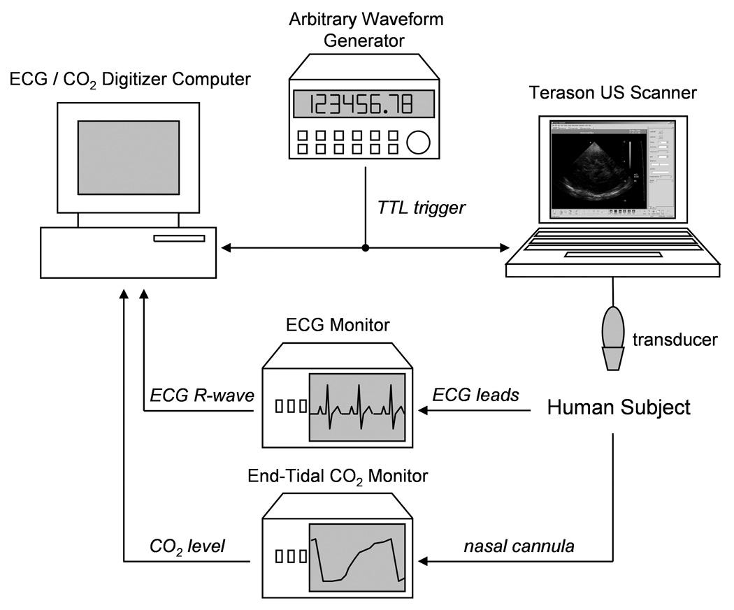
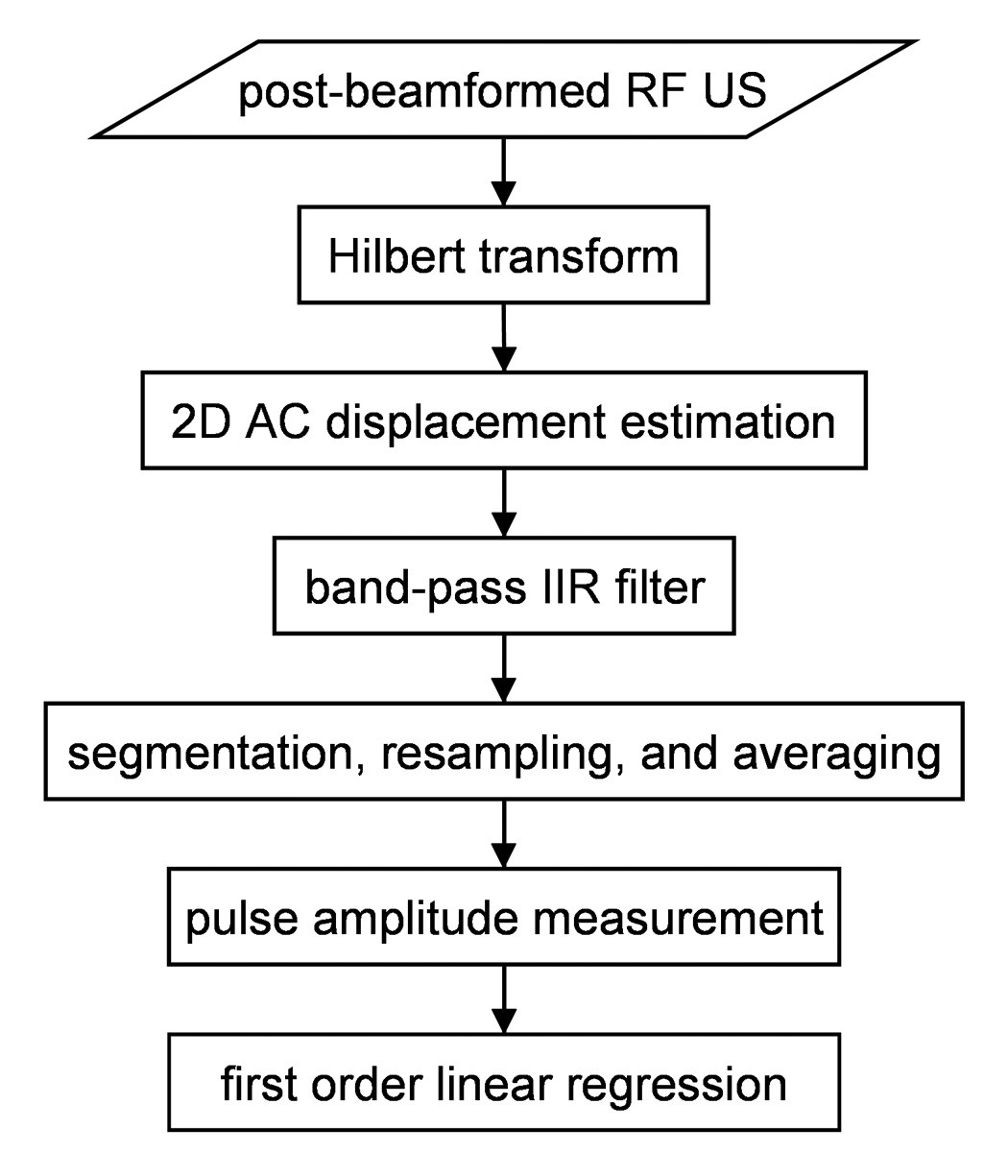
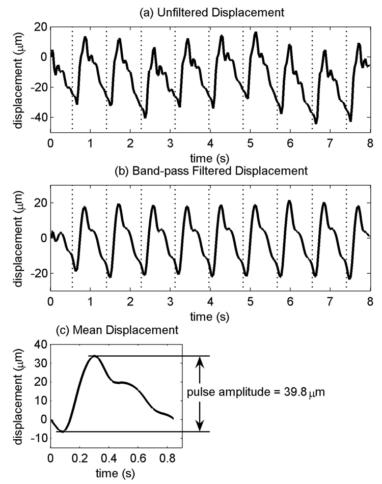
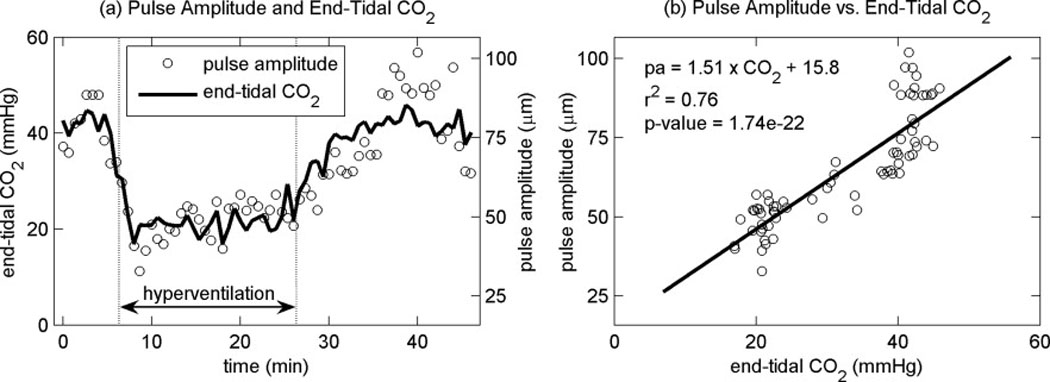
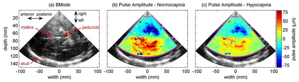
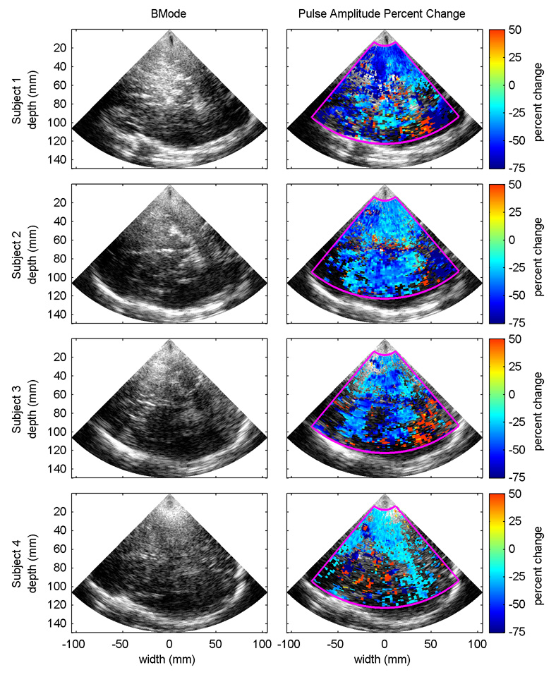
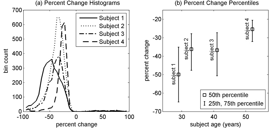
Tissue pulsatility imaging (TPI) is an ultrasonic technique that is being developed at the University of Washington to measure tissue displacement or strain as a result of blood flow over the cardiac and respiratory cycles. This technique is based in principle on plethysmography, an older nonultrasound technology for measuring expansion of a whole limb or body part due to perfusion. TPI adapts tissue Doppler signal processing methods to measure the "plethysmographic" signal from hundreds or thousands of sample volumes in an ultrasound image plane. This paper presents a feasibility study to determine if TPI can be used to assess cerebral vasoreactivity. Ultrasound data were collected transcranially through the temporal acoustic window from four subjects before, during and after voluntary hyperventilation. In each subject, decreases in tissue pulsatility during hyperventilation were observed that were statistically correlated with the subject's end-tidal CO2 measurements. (
Figures
References
-
- Akin A, Bilensoy D. Cerebrovascular reactivity to hypercapnia in migraine patients measured with near-infrared spectroscopy. Brain Res. 2006;1107(1):206–214. - PubMed
-
- Beach KW, Philips DJ, Kansky J. Ultrasonic plethysmograph. US Patent. # 5,088,498. 1992.
-
- Beach KW, Philips DJ, Kansky J. Ultrasonic plethysmograph. US Patent. # 5,183,046. 1993.
-
- Beach KW, Philips DJ, Kansky J. Ultrasonic plethysmograph. US Patent. # 5,289,820. 1994.
-
- de Boorder MJ, van der Grond J, van Dongen AJ, Klijn CJ, Jaap Kappelle L, Van Rijk PP, Hendrikse J. Spect measurements of regional cerebral perfusion and carbondioxide reactivity: correlation with cerebral collaterals in internal carotid artery occlousive disease. J Neurol. 2006;253(10):1285–1291. - PubMed
Publication types
MeSH terms
Grants and funding
LinkOut - more resources
Full Text Sources
Other Literature Sources
Miscellaneous
