

Acupuncture modulates resting state connectivity in default and sensorimotor brain networks
- PMID: 18337009
- PMCID: PMC2440647
- DOI: 10.1016/j.pain.2008.01.011
Acupuncture modulates resting state connectivity in default and sensorimotor brain networks
Abstract
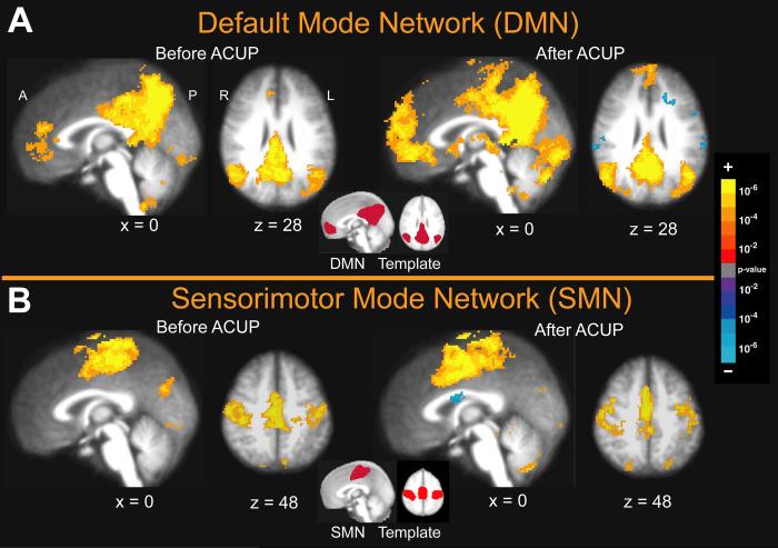
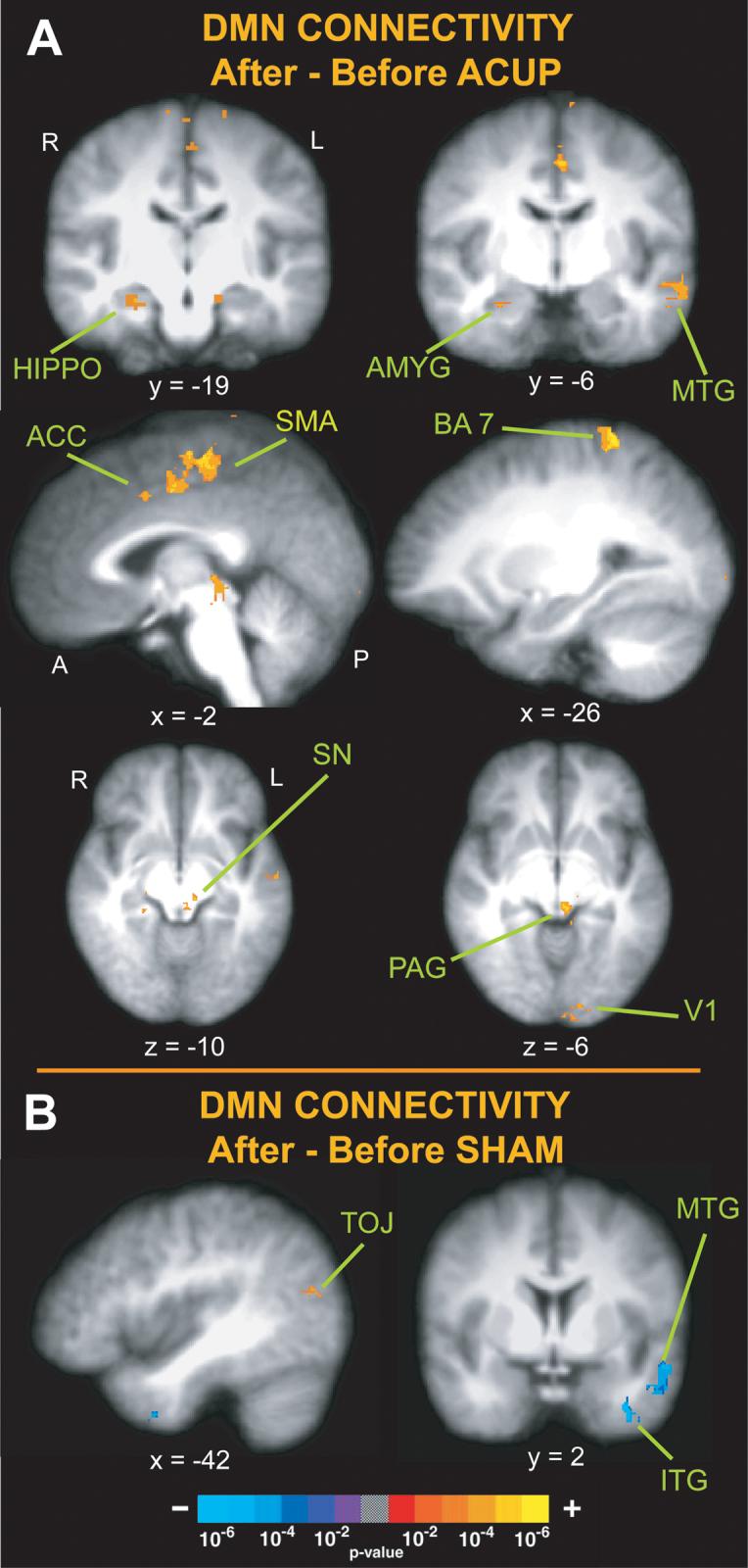
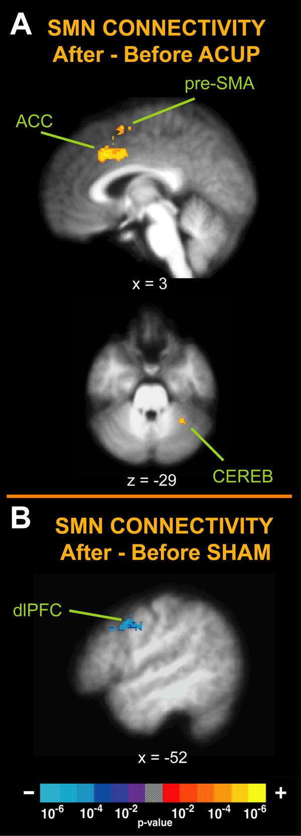
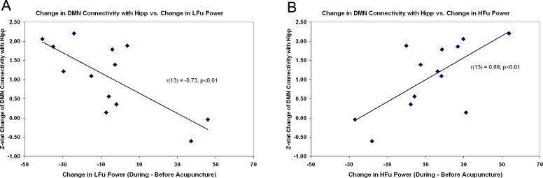
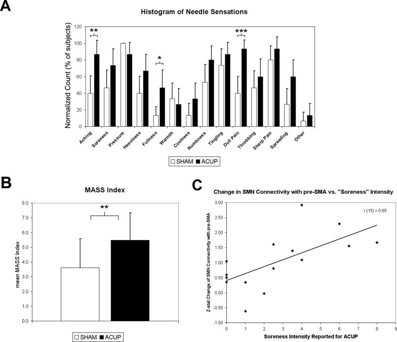
Previous studies have defined low-frequency, spatially consistent networks in resting fMRI data which may reflect functional connectivity. We sought to explore how a complex somatosensory stimulation, acupuncture, influences intrinsic connectivity in two of these networks: the default mode network (DMN) and sensorimotor network (SMN). We analyzed resting fMRI data taken before and after verum and sham acupuncture. Electrocardiography data were used to infer autonomic modulation through measures of heart rate variability (HRV). Probabilistic independent component analysis was used to separate resting fMRI data into DMN and SMN components. Following verum, but not sham, acupuncture there was increased DMN connectivity with pain (anterior cingulate cortex (ACC), periaqueductal gray), affective (amygdala, ACC), and memory (hippocampal formation, middle temporal gyrus) related brain regions. Furthermore, increased DMN connectivity with the hippocampal formation, a region known to support memory and interconnected with autonomic brain regions, was negatively correlated with acupuncture-induced increase in a sympathetic related HRV metric (LFu), and positively correlated with a parasympathetic related metric (HFu). Following verum, but not sham, acupuncture there was also increased SMN connectivity with pain-related brain regions (ACC, cerebellum). We attribute differences between verum and sham acupuncture to more varied and stronger sensations evoked by verum acupuncture. Our results demonstrate for the first time that acupuncture can enhance the post-stimulation spatial extent of resting brain networks to include anti-nociceptive, memory, and affective brain regions. This modulation and sympathovagal response may relate to acupuncture analgesia and other potential therapeutic effects.
Figures
Comment in
-
Acupuncture and the CNS: what can the brain at rest suggest?Pain. 2008 Jun;136(3):230-231. doi: 10.1016/j.pain.2008.03.008. Epub 2008 Apr 18. Pain. 2008. PMID: 18423873 No abstract available.
References
-
- Andersson S, Lundeberg T. Acupuncture - from empiricism to science: functional background to acupuncture effects in pain and disease. Medical Hypotheses. 1995;45:271–281. - PubMed
-
- Baliki M, Geha P, Apkarian A, Chialvo D. Society for Neuroscience Annual Meeting. San Diego: 2007. Impaired brain de-activation in chronic pain.
-
- Bantick SJ, Wise RG, Ploghaus A, Clare S, Smith SM, Tracey I. Imaging how attention modulates pain in humans using functional MRI. Brain. 2002;125(Pt 2):310–9. - PubMed
-
- Becerra L, Breiter HC, Wise R, Gonzalez RG, Borsook D. Reward circuitry activation by noxious thermal stimuli. Neuron. 2001;32(5):927–46. - PubMed
-
- Becerra LR, Breiter HC, Stojanovic M, Fishman S, Edwards A, Comite AR, Gonzalez RG, Borsook D. Human brain activation under controlled thermal stimulation and habituation to noxious heat: an fMRI study. Magn Reson Med. 1999;41(5):1044–57. - PubMed
