. 2008 May;127(2):176-87.
doi: 10.1016/j.clim.2007.12.009. Epub 2008 Mar 12.

Tissue-targeted therapy of autoimmune diabetes using dendritic cells transduced to express IL-4 in NOD mice

Affiliations

Tissue-targeted therapy of autoimmune diabetes using dendritic cells transduced to express IL-4 in NOD mice

Rémi J Creusot et al. Clin Immunol. 2008 May.

Abstract

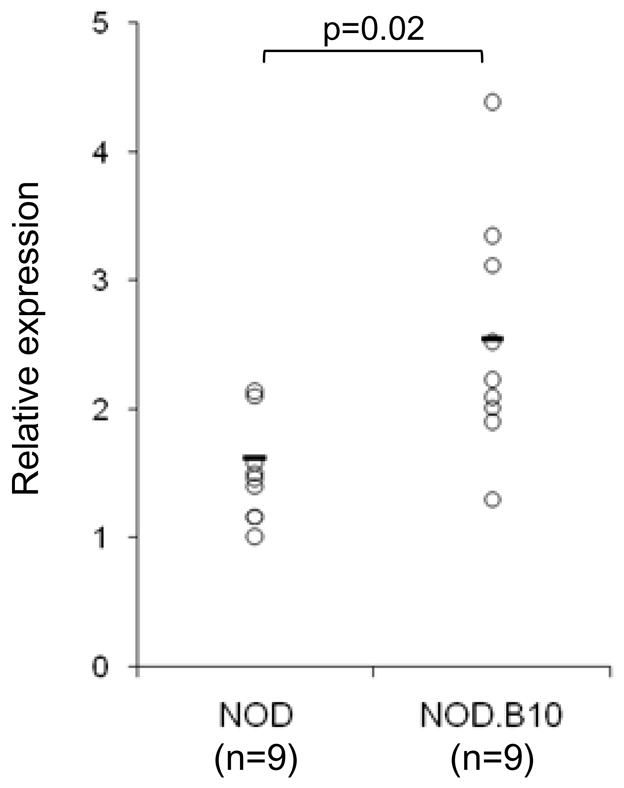
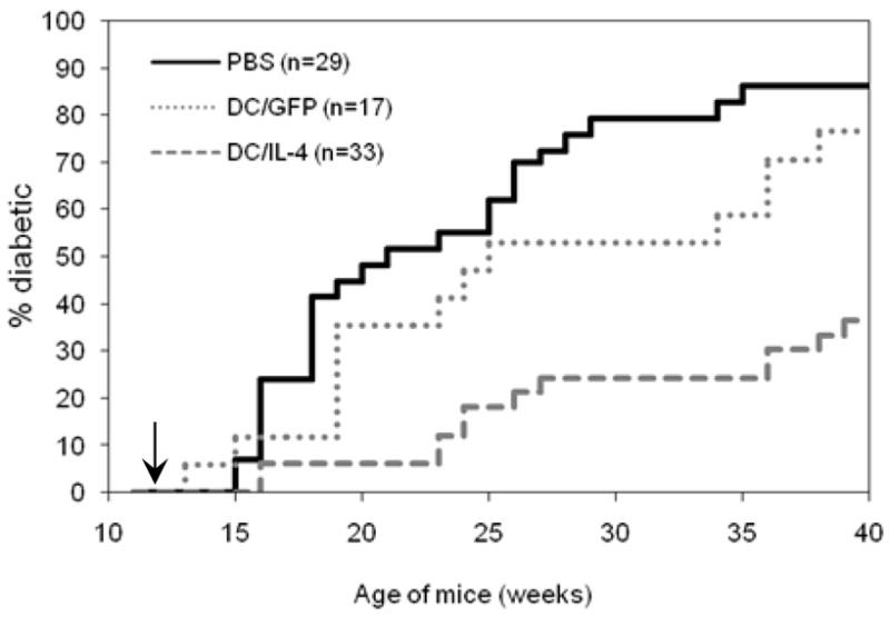
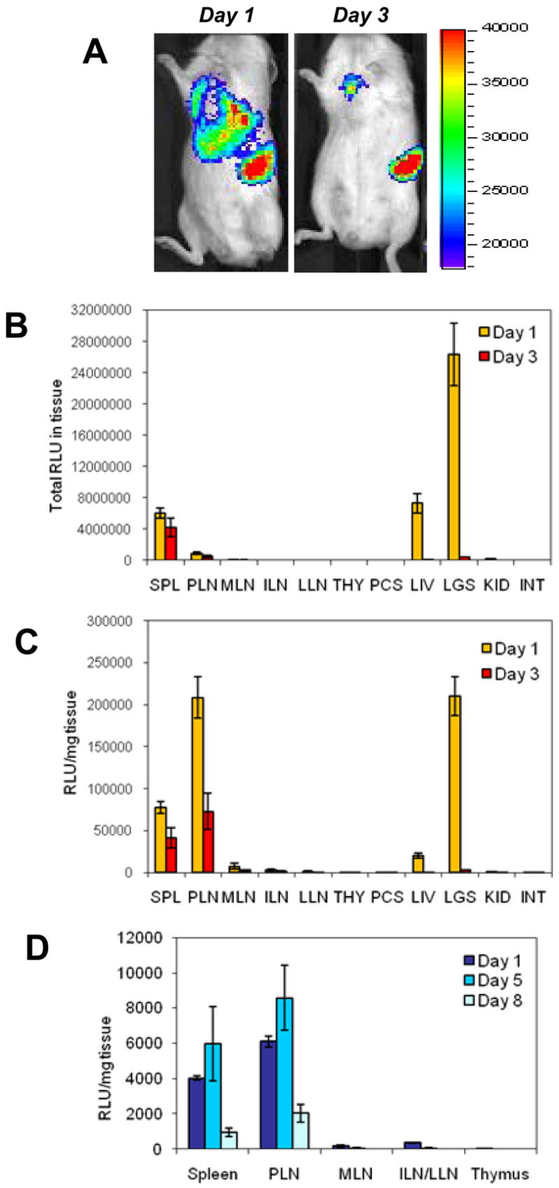
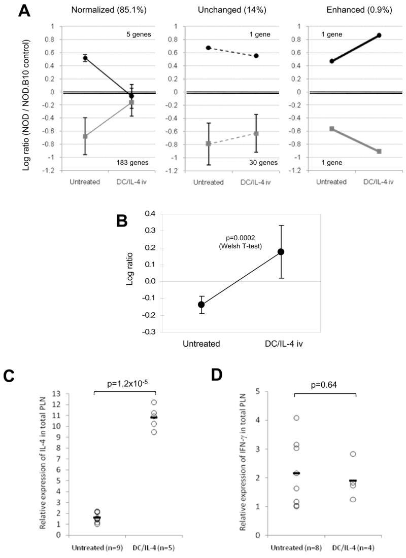
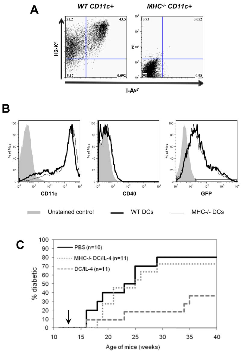
A deficit in IL-4 production has been previously reported in both diabetic human patients and non-obese diabetic (NOD) mice. In addition, re-introducing IL-4 into NOD mice systemically, or as a transgene, led to a beneficial outcome in most studies. Here, we show that prediabetic, 12-week old female NOD mice have a deficit in IL-4 expression in the pancreatic lymph nodes (PLN) compared to age-matched diabetes-resistant NOD.B10 mice. By bioluminescence imaging, we demonstrated that the PLN was preferentially targeted by bone marrow-derived dendritic cells (DCs) following intravenous (IV) administration. Following IV injection of DCs transduced to express IL-4 (DC/IL-4) into 12-week old NOD mice, it was possible to significantly delay or prevent the onset of hyperglycemia. We then focused on the PLN to monitor, by microarray analysis, changes in gene expression induced by DC/IL-4 and observed a rapid normalization of the expression of many genes, that were otherwise under-expressed compared to NOD.B10 PLN. The protective effect of DC/IL-4 required both MHC and IL-4 expression by the DCs. Thus, adoptive cellular therapy, using DCs modified to express IL-4, offers an effective, tissue-targeted cellular therapy to prevent diabetes in NOD mice at an advanced stage of pre-diabetes, and may offer a safe approach to consider for treatment of high risk human pre-diabetic patients.


Figures

Figure 1
Figure 1
IL-4 deficiency in the PLN of 12-wk old NOD mice. IL-4 expression in the PLN of 12-wk old euglycemic NOD (n=9) and NOD.B10 (n=9) was determined by RT-PCR. The expression was normalized to β actin. Individual values and mean are shown and T-test was applied for statistical analysis.
Figure 2
Figure 2
Characterization of DCs used for adoptive cellular gene therapy. Untransduced DCs (DC/Unt) and transduced DCs (DC/GLF and DC/IL-4) were analyzed by flow cytometry. Plots were all gated on live DCs based on FSc and PI profile. (A) Purity was determined based on CD11c and CD11b expression. (B) Transduction efficiency was assessed by GFP expression on live DCs. Surface marker levels (CD11c, CD40, CD80, CD86 and class II MHC I-Ag7) were analyzed on total live DCs for DC/Unt (grey-filled histograms), or on the GFP+ live DC fraction for DC/GLF (dashed line) and DC/IL-4 (thick line). (C) After harvest on day 6, transduced DCs (DC/GFP and DC/IL-4) were re-plated at various cell numbers in cytokine-free medium for another 36h, after which supernatant was collected and used for ELISA (one of two representative experiments shown).
Figure 3
Figure 3
Incidence of diabetes after IV treatment of 12-wk old prediabetic female NOD mice with PBS, DC/GFP or DC/IL-4. Combined results from 3 similar, independent experiments (5–7.5x10⁵ GFP+ DCs/mouse) are shown. Statistical analysis: PBS vs DC/IL-4, p=2x10⁻⁶ by Log Rank and 5x10⁻⁶ by Wilcoxon; PBS vs DC/GFP, p=0.18 by Log Rank and Wilcoxon; DC/GFP vs DC/IL-4, p=0.002 by Log Rank and Wilcoxon.
Figure 4
Figure 4
In vivo bioluminescence imaging (A) and biodistribution analysis (B, C) of Luc+ NOD DCs, 1 and 3 days after IV injection. DC/GLF (1x10⁶ GFP+ cells) were injected into 12-wk old euglycemic NOD mice. Mice were imaged live at various time points (A), and some of them sacrificed for biodistribution analysis. Total luciferase activity in tissues (B) and normalized activity per mg tissue (C) are shown (mean ± SE, n=3 mice per time point, one of two similar experiments shown). Migration of Luc+ NOD.B10 DCs after IV injection (5x10⁵ GFP+ cells/mouse) into 12-wk old NOD.B10 mice was also evaluated. Panel (D) shows normalized luciferase activity per mg tissue at different time points after IV injection (mean ± SE, n=3 mice per time point). Abbreviations: SPL, spleen; PLN, pancreatic LNs; MLN, mesenteric LNs; ILN, inguinal LNs; LLN, lumbar LNs; THY, thymus; PCS, pancreas; LIV, liver (left lobe only); LGS, lungs; KID, kidney; INT, ileum part of intestine.
Figure 5
Figure 5
Changes in gene expression induced by DC/IL-4 in the PLN of 12 wk-old NOD mice. (A) Gene expression in PLN from female NOD mice, untreated (n=7) or treated with DC/IL-4 iv (n=9), was analyzed against a tissue-matched control from female NOD.B10 mice. A total of 221 genes, which expression was ~3 fold over- (log10 ratio >0.45) or under-expressed (log10 ratio <-0.45) in NOD compared to NOD.B10 (p<0.01) were selected. Their change, 3 days following treatment with DC/IL-4, was graphically represented in three groups (normalized, unchanged or enhanced; mean of log10 ratios ± SD) using data shown on Table 1. Normalized or enhanced gene expression were significantly changed by DC/IL-4 treatment (p<0.05). (B) IL-4 expression in NOD PLN, relative to NOD.B10 control, before and after DC/IL-4 treatment (mean ± SD; p=0.0002, T-test). IL-4 was not part of the above genes, because although significantly under-expressed, its log10 ratio was <0.45. (C, D) Relative expression of IL-4 (C) and IFN-γ (D) by RT-PCR in PLN of untreated or DC/IL-4-treated 12-wk old NOD mice (individual results and mean; data normalized to β actin; T-test applied).
Figure 6
Figure 6
MHC expression is required for protection in addition to IL-4 secretion. (A) Class I and class II MHC expression on DCs from normal or MHC−/− NOD mice. (B) Similar expression of CD11c, CD40 and transduction efficiency in DCs from normal or MHC−/− NOD mice. (C) Incidence of diabetes after IV treatment of 12-wk old prediabetic female NOD mice with either PBS, DC/IL-4 or MHC−/− DC/IL-4 (7.5x10⁵ GFP+ DCs/mouse). Statistical analysis (Log Rank test): PBS vs DC/IL-4, p=0.02; PBS vs MHC−/− DC/IL-4, p=0.6; MHC−/− DC/IL-4 vs DC/IL-4, p=0.05.

Similar articles

Cited by

References

    1. Shoda L, Young D, Ramanujan S, Whiting C, Atkinson M, Bluestone J, Eisenbarth G, Mathis D, Rossini A, Campbell S, Kahn R, Kreuwel H. A comprehensive review of interventions in the NOD mouse and implications for translation. Immunity. 2005;23:115–26. - PubMed
    1. Staeva-Vieira T, Peakman M, von Herrath M. Translational mini-review series on type 1 diabetes: Immune-based therapeutic approaches for type 1 diabetes. Clin Exp Immunol. 2007;148:17–31. - PMC - PubMed
    1. Roep B, Atkinson M, von Herrath M. Satisfaction (not) guaranteed: re-evaluating the use of animal models of type 1 diabetes. Nat Rev Immunol. 2004;4:989–97. - PubMed
    1. Creusot R, Fathman CG, Muller-Ladner U, Tarner I. Targeted gene therapy of autoimmune diseases: advances and prospects. Expert Rev Clin Immunol. 2005;1:385–404. - PubMed
    1. Cameron M, Arreaza G, Zucker P, Chensue S, Strieter R, Chakrabarti S, Delovitch T. IL-4 prevents insulitis and insulin-dependent diabetes mellitus in nonobese diabetic mice by potentiation of regulatory T helper-2 cell function. J Immunol. 1997;159:4686–92. - PubMed

Publication types

MeSH terms