. 2008 May 9;283(19):13044-52.
doi: 10.1074/jbc.M800900200. Epub 2008 Mar 12.

The cytoplasmic loops of subunit a of Escherichia coli ATP synthase may participate in the proton translocating mechanism

Affiliations

The cytoplasmic loops of subunit a of Escherichia coli ATP synthase may participate in the proton translocating mechanism

Kyle J Moore et al. J Biol Chem. .

Abstract

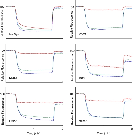
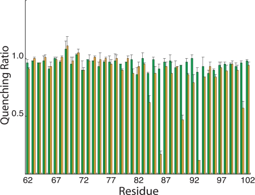
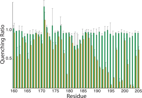
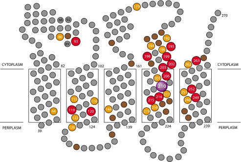
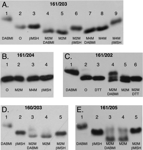
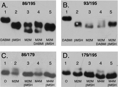
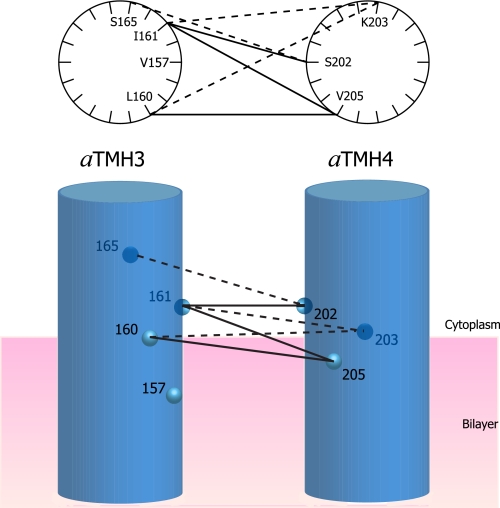
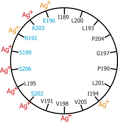
Subunit a plays a key role in promoting H(+) transport and the coupled rotary motion of the subunit c ring in F(1)F(0)-ATP synthase. H(+) binding and release occur at Asp-61 in the middle of the second transmembrane helix (TMH) of F(0) subunit c. H(+) are thought to reach Asp-61 via aqueous pathways mapping to the surfaces of TMHs 2-5 of subunit a based upon the chemical reactivity of Cys substituted into these helices. Here we substituted Cys into loops connecting TMHs 1 and 2 (loop 1-2) and TMHs 3 and 4 (loop 3-4). A large segment of loop 3-4 extending from loop residue 192 loop to residue 203 in TMH4 at the lipid bilayer surface proved to be very sensitive to inhibition by Ag(+). Cys-161 and -165 at the other end of the loop bordering TMH3 were also sensitive to inhibition by Ag(+). Further Cys substitutions in residues 86 and 93 in the middle of the 1-2 loop proved to be Ag(+)-sensitive. We next asked whether the regions of Ag(+)-sensitive residues clustered together near the surface of the membrane by combining Cys substitutions from two domains and testing for cross-linking. Cys-161 and -165 in loop 3-4 were found to cross-link with Cys-202, -203, or -205, which extend into TMH4 from the cytoplasm. Further Cys at residues 86 and 93 in loop 1-2 were found to cross-link with Cys-195 in loop 3-4. We conclude that the Ag(+)-sensitive regions of loops 1-2 and 3-4 may pack in a single domain that packs at the ends of TMHs 3 and 4. We suggest that the Ag(+)-sensitive domain may be involved in gating H(+) release at the cytoplasmic side of the aqueous access channel extending through F(0).


Figures

FIGURE 1.
FIGURE 1.
Differing sensitivity of Cys substitutions in 1–2 loop and 3–4 loop to Ag+ and NEM inhibition. A 160-μl aliquot of membranes at 10 mg/ml in TMG-acetate were treated with 5 mm NEM for 15 min at room temperature and then diluted into 3.2 ml of HMK-nitrate buffer containing 0.3 μg/ml ACMA. Alternatively membranes were diluted into HMK-nitrate buffer, and AgNO3 was added to 40 μm for 15 min at room temperature prior to addition of ACMA. ATP was added to 0.94 mm at 20 s, and the uncoupler nigericin was added to 0.5 μg/ml at 100 s. The traces indicate no treatment (blue), NEM treatment (green), or Ag+ treatment (red). The substitution tested is indicated in each panel.
FIGURE 2.
FIGURE 2.
NEM and Ag+ sensitivity of Cys substitutions in the 1–2 loop. Results are presented as the ratios of the quenching response in the presence of Ag+ or NEM to the quenching response in the absence of either reagent. The green bars represent the quenching ratio ±NEM treatment, whereas the orange bars represent the quenching ratio ±Ag+ treatment. Each bar represents the average ratio from n ≥ 2 determinations ±S.D.
FIGURE 3.
FIGURE 3.
NEM and Ag+ sensitivity of Cys substitutions in the 3–4 loop. Results are presented as the ratios of the quenching response in the presence of Ag+ or NEM to the quenching response in the absence of either reagent. The green bars represent the quenching ratio ±NEM treatment, whereas the orange bars represent the quenching ratio ±Ag+ treatment. Each bar represents the average ratio from n ≥ 2 determinations ±S.D.
FIGURE 4.
FIGURE 4.
Position of Ag+-sensitive residues in a two-dimensional topological map of subunit a. The suggested regions of α-helical secondary structure depicted in the 1–2 and 3–4 loops are based upon NMR chemical shift analysis of subunit a in chloroform-methanol-H2O (4:4:1) solvent (53). Residues that are most sensitive to inhibition by Ag+ are shown in red (>85% inhibition), orange (66–85% inhibition), and brown (46–65% inhibition). The relative depth of placement of the five TMHs of subunit a in the lipid bilayer is based upon cross-linking experiments as described elsewhere (19).
FIGURE 5.
FIGURE 5.
Cross-link formation between pairs of Cys substitutions placed in the two ends of the 3–4 loop. Membranes from the double Cys substitutions shown were treated with the reagents shown in each lane in the sequence indicated as described under “Experimental Procedures.” Following SDS-PAGE, Western blots were probed with anti-subunit a antibody. Cross-link formation led to increased electrophoretic mobility and a downward band shift as discussed in the text. A, I161C/K203C membranes; B, I161C/204 membranes; C, I161C/P204C membranes; D, L160C/K203C membranes; E, I161C/V205C membranes. βMSH, β-mercaptoethanol; DTT, dithiothreitol; O, no treatment.
FIGURE 6.
FIGURE 6.
Cross-link formation between Cys substitutions in the center of the 1–2 loop and C-terminal end of the 3–4 loop. Membranes from the double Cys substitutions were treated with the reagents shown in each lane in the sequence indicated as described under “Experimental Procedures.” Following SDS-PAGE, Western blots were probed with anti-subunit a antibody. Cross-link formation led to increased electrophoretic mobility and a downward band shift as discussed in the text. A, V86C/L195C membranes; B, M93C/L195C membranes; C, V86C/T179C membranes; D, T179C/L195C membranes. βMSH, β-mercaptoethanol; O, no treatment.
FIGURE 7.
FIGURE 7.
Cross-link formation between modeled α-helical faces of aTMH3 and aTMH4 as they emerge from the cytoplasmic face of the membrane. The double Cys pairs that cross-link with M2M are tabulated in Table 2 and here shown connected by lines. The light or dark circles are shown to distinguish the front and back faces of the cylinders. The solid versus dashed lines are intended to distinguish cross-linking to the front and back faces of the α-helical wheels or cylinders. The M4M cross-link between 157 and 203 is not connected by a line.
FIGURE 8.
FIGURE 8.
Amphipathic characteristics and Ag+ sensitivity of residues in an α-helical wheel model for the C-terminal region of the 3–4 loop. Polar or charged wild type residues are shown in blue, and hydrophobic residues are shown in black. Ag+-sensitive Cys substitutions are highlighted with red Ag+ indicating >85% inhibition and orange Ag+ indicating 66–85% inhibition.

References

    1. Yoshida, M., Muneyuki, E., and Hisabori, T. (2001) Nat. Rev. Mol. Biol. 2 669–677 - PubMed
    1. Capaldi, R. A., and Aggeler, R. (2002) Trends Biochem. Sci. 27 154–160 - PubMed
    1. Dimroth, P., von Ballmoos, C., and Meier, T. (2006) EMBO Rep. 7 276–282 - PMC - PubMed
    1. Senior, A. E. (1988) Physiol. Rev. 68 177–231 - PubMed
    1. Jiang, W., Hermolin, J., and Fillingame, R. H. (2001) Proc. Natl. Acad. Sci. U. S. A. 98 4966–4971 - PMC - PubMed

Publication types

MeSH terms

LinkOut - more resources