. 2008 May 16;283(20):14072-83.
doi: 10.1074/jbc.M708551200. Epub 2008 Mar 12.

Activated nuclear metabotropic glutamate receptor mGlu5 couples to nuclear Gq/11 proteins to generate inositol 1,4,5-trisphosphate-mediated nuclear Ca2+ release

Affiliations

Activated nuclear metabotropic glutamate receptor mGlu5 couples to nuclear Gq/11 proteins to generate inositol 1,4,5-trisphosphate-mediated nuclear Ca2+ release

Vikas Kumar et al. J Biol Chem. .

Abstract

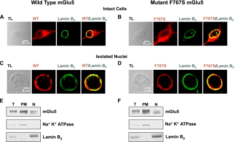
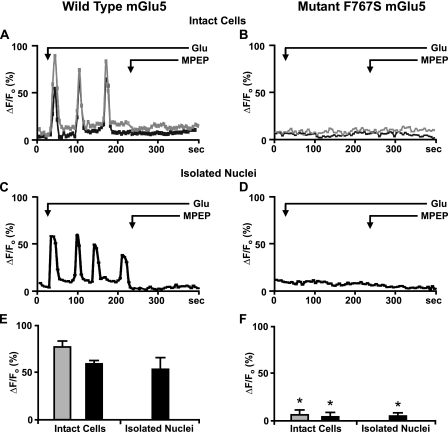
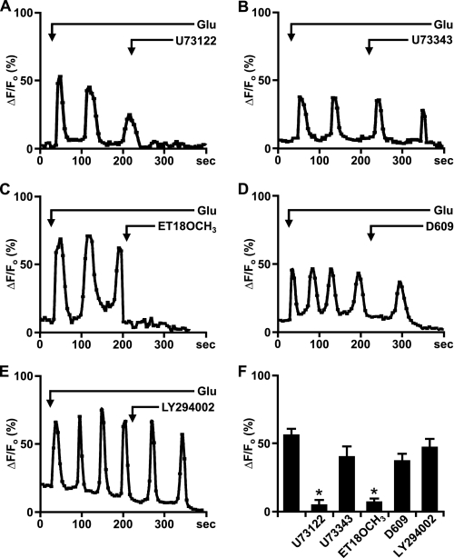
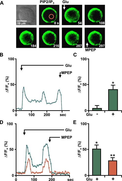
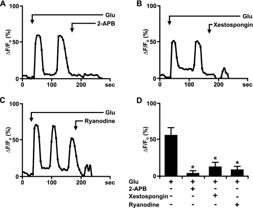
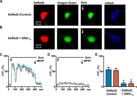
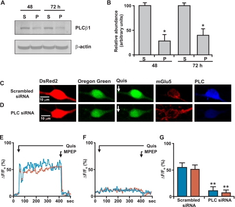
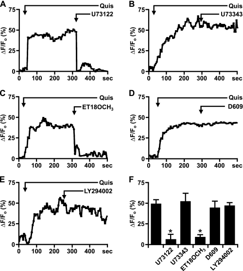
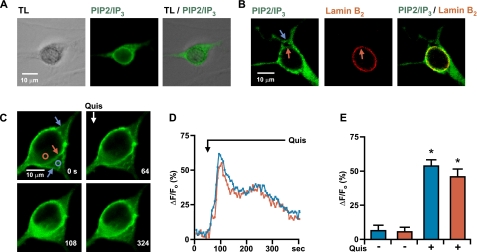
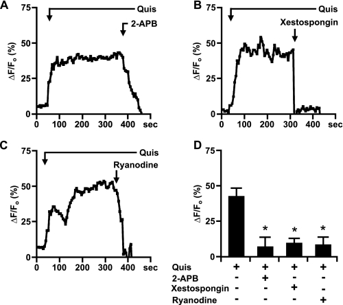
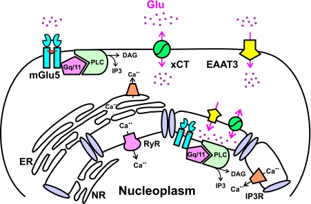
Recently we have shown that the metabotropic glutamate 5 (mGlu5) receptor can be expressed on nuclear membranes of heterologous cells or endogenously on striatal neurons where it can mediate nuclear Ca2+ changes. Here, pharmacological, optical, and genetic techniques were used to show that upon activation, nuclear mGlu5 receptors generate nuclear inositol 1,4,5-trisphosphate (IP3) in situ. Specifically, expression of an mGlu5 F767S mutant in HEK293 cells that blocks Gq/11 coupling or introduction of a dominant negative Galphaq construct in striatal neurons prevented nuclear Ca2+ changes following receptor activation. These data indicate that nuclear mGlu5 receptors couple to Gq/11 to mobilize nuclear Ca2+. Nuclear mGlu5-mediated Ca2+ responses could also be blocked by the phospholipase C (PLC) inhibitor, U73122, the phosphatidylinositol (PI) PLC inhibitor 1-O-octadecyl-2-O-methyl-sn-glycero-3-phosphorylcholine (ET-18-OCH3), or by using small interfering RNA targeted against PLCbeta1 demonstrating that PI-PLC is involved. Direct assessment of inositol phosphate production using a PIP2/IP3 "biosensor" revealed for the first time that IP3 can be generated in the nucleus following activation of nuclear mGlu5 receptors. Finally, both IP3 and ryanodine receptor blockers prevented nuclear mGlu5-mediated increases in intranuclear Ca2+. Collectively, this study shows that like plasma membrane receptors, activated nuclear mGlu5 receptors couple to Gq/11 and PLC to generate IP3-mediated release of Ca2+ from Ca2+-release channels in the nucleus. Thus the nucleus can function as an autonomous organelle independent of signals originating in the cytoplasm, and nuclear mGlu5 receptors play a dynamic role in mobilizing Ca2+ in a specific, localized fashion.


Figures

FIGURE 1.
FIGURE 1.
mGlu5 wild type and F767S mutant exhibit nuclear localization. Stable HEK cell lines expressing either wild type mGlu5 or the F767S mutant, were fixed, permeabilized, and processed for immunocytochemistry using receptor-specific antibodies as well as anti-lamin B2. Cells were analyzed by confocal microscopy to detect receptor localization (red) or lamin B2 distribution (green). Photographs represent single optical sections of 0.4 μm merged such that yellow indicates co-localization of the specific antigens. Receptors expressed by intact cells (A and B; TL, transmitted light) or their isolated nuclei (C and D) exhibited pronounced co-localization with lamin B2. Subcellular fractionation of HEK cell lines expressing either wild type mGlu5 (E) or the F767S mutant (F) shows that wild type or mutant receptors can be detected in fractions containing either the nuclear (N) or plasma membranes (PM). Equal amounts of protein (30 μg) from each fraction were separated on reducing SDS gels and transferred to nylon membranes (T, total cell lysates). The same blot was sequentially probed with antibodies against mGlu5, the inner nuclear marker, lamin B2, and the plasma membrane marker, Na+K+-ATPase.
FIGURE 2.
FIGURE 2.
Nuclear mGlu5 couples to nuclear Gq/11 protein. Representative traces are shown of cytoplasmic (gray line) or nuclear (black line) Ca2+ responses from wild type mGlu5 expressing HEK cells (A) or the F767S cell line (B) as well as their respective nuclei (C and D) following bath addition of 10 μm glutamate (Glu) and 1 μm MPEP when indicated and scanned at 5.4 s/scan. Oscillations are represented as the fractional change in fluorescence relative to the basal value. Compiled data from the maximum response of initial peak (ΔF/Fo, %) from either wild type cells or nuclei (E) or F767S cells or nuclei (F) from n > 30 cells, from three to five independent experiments. *, p < 10-4 when compared with Ca2+ responses from wild type mGlu5/HEK cells and nuclei treated with glutamate only.
FIGURE 3.
FIGURE 3.
Nuclear mGlu5 stimulates nuclear PI-PLC. A-E, representative traces of nuclear (black line) Ca2+ responses in isolated mGlu5-expressing HEK nuclei represented as the fractional change in fluorescence relative to the basal level. Isolated nuclei were treated with 10 μm glutamate and the specified antagonists bath applied when indicated by the arrow; U73122 (PLC inhibitor, 3 μm), U73343 (inactive analog of U73122, 3 μm), ET-18-OCH3 (selective PI-PLC inhibitor, 10 μm), D609 (selective PC-PLC inhibitor, 100 μm), and LY294002 (PI3K inhibitor, 100 nm). F, compiled data from maximum response of initial peak (ΔF/Fo, %) induced by glutamate alone or together with specified antagonists from n > 15 in all cases, from three independent experiments. *, p < 0.001 when compared with Ca2+ responses from nuclei treated with glutamate only.
FIGURE 4.
FIGURE 4.
Nuclear mGlu5-generated IP3 production can be measured using PIP2/IP3 biosensor. A, first panel, transmitted light image of selected nucleus; remaining panels, time lapse imaging of the mGlu5/HEK nucleus transiently transfected with the PIP2/IP3 biosensor treated at the indicated times (seconds) with 10 μm glutamate or 1 μm MPEP. Red circle corresponds to area measured for representative trace shown in B, where Glu-mediated oscillations are represented as the fractional change in fluorescence relative to the basal IP3 levels. C, compiled data from maximum response of the initial peak (ΔF/Fo, %) for nuclear responses from n = 5, from three experiments. *, p < 0.001 when compared with baseline IP3 responses. D, nuclei isolated from a mGlu5/HEK stable cell line transiently transfected with the PIP2/IP3 biosensor were loaded with Calcium Crimson-AM to simultaneously measure Ca2+ and biosensor changes. Shown is a representative trace of nuclear Ca2+ (red line) and PIP2/IP3 biosensor (green line) responses in an isolated nucleus treated with 10 μm glutamate and 1 μm MPEP as indicated by the arrow. E, compiled data from the maximum response of initial peak (ΔF/Fo, %) for nuclear IP3 (green) and Ca2+ (red) responses from n = 5, from three independent experiments. *, p < 0.001 when compared with baseline IP3 response; **, p < 0.01 when compared with the baseline Ca2+ response.
FIGURE 5.
FIGURE 5.
Glutamate-activated nuclear mGlu5-mediated Ca2+ changes can be abrogated by IP3R and RyR blockers. A-C, representative traces of nuclear (black line) Ca2+ responses in isolated mGlu5-expressing nuclei represented as the fractional change in fluorescence relative to the basal level. Isolated nuclei were treated with 10 μm glutamate and the indicated antagonists; 2-APB (IP3R modulator, 100 μm), xestospongin C (IP3R antagonist, 2 μm), and ryanodine (RyR antagonist, 100 μm). D, compiled data from the maximum response of the initial peak (ΔF/Fo, %) for nuclear responses from n > 20 for 2-APB and ryanodine and n = 14 for xestospongin C, from three independent experiments. *, p < 0.001 when compared with Ca2+ responses from nuclei treated with glutamate only.
FIGURE 6.
FIGURE 6.
Endogenous mGlu5 receptors expressed on striatal neurons couple to the Gq family of G-proteins. On the 12th day in vitro, striatal neurons transiently transfected with DsRed2 (A) or co-transfected with dominant negative Gαq and DsRed2 (B) were loaded with Oregon Green BAPTA-1AM (second panels) and quisqualate (Quis;10 μm) was bath applied (third panels). Remaining panels, images of post hoc identified mGlu5-positive cells. Representative traces are shown of quisqualate-mediated cytoplasmic (blue line) or nuclear (red line) Ca2+ responses from a control neuron (DsRed2 transfected; C) or a neuron co-transfected with dominant negative Gαq and DsRed2 (D). MPEP was added as indicated (1 μm; black line). E, compiled data from the maximum response of initial peak (ΔF/Fo, %) from either control cells (n = 22) or dominant negative Gαq-transfected neurons (n = 20) from four independent experiments. *, p < 0.0001 when compared with Ca2+ responses from DsRed2-transfected control cells.
FIGURE 7.
FIGURE 7.
Knockdown of PLCβ1 leads to reduction in striatal mGlu5-mediated Ca2+ changes. A, primary striatal neurons were transiently transfected with either scrambled siRNA (S) or PLCβ1 siRNA (P). At the times indicated, cells were lysed and proteins were separated on reducing SDS gels and transferred to nylon membranes. The same blot was sequentially probed with antibodies against PLCβ1 (Santa Cruz) and β-actin. B, the relative abundance of PLCβ1 was measured by Western blotting; the efficiency of knockdown is expressed in arbitrary units compared with the PLCβ1 levels in scrambled siRNA-transfected neurons. The data shown are compiled from three independent experiments. *, p < 0.005 when compared with PLCβ1 protein levels in scrambled siRNA-transfected cells. On the 12th day in vitro, striatal neurons were transiently co-transfected with scrambled siRNA and DsRed2 (C) or with PLCβ1 siRNA and DsRed2 (D). Two days later, neurons were loaded with Oregon Green BAPTA-1AM (second panels) and quisqualate (Quis;10 μm) was bath applied (third panels). Representative traces are shown for cytoplasmic (blue line) or nuclear (red line) Ca2+ responses from scrambled (E) or PLCβ1 siRNA (F) following quisqualate and subsequently, 1 μm MPEP administration (black line). Imaged neurons were post hoc identified using mGlu5 (fourth panels) or PLCβ1 antibodies (last panels). G, compiled data from the maximum response of initial peak (ΔF/Fo, %) from either scrambled (n = 13) or PLCβ1 (n = 17) siRNA-transfected neurons from three independent experiments. **, p < 0.0001 when compared with Ca2+ responses from control siRNA and DsRed2-transfected cells.
FIGURE 8.
FIGURE 8.
Endogenous mGlu5 receptors expressed on striatal nuclei mediate Ca2+ changes via the PI-PLC pathway. P10 striatal nuclei were acutely isolated, loaded with Oregon Green BAPTA-1AM, and imaged in real time. A-E, quisqualate-mediated (10 μm) representative traces of nuclear (black line) Ca2+ responses treated with the indicated antagonists bath applied as noted. F, compiled data from the maximum response of initial peak (ΔF/Fo, %) for nuclear Ca2+ responses induced by quisqualate alone or together with specified antagonists from n > 8 in all cases, from three to four independent experiments. *, p < 0.001 when compared with Ca2+ responses from nuclei treated only with quisqualate (Quis).
FIGURE 9.
FIGURE 9.
Activation of striatal mGlu5 receptors generates nuclear IP3. On the 12th day in vitro, striatal cultures were transiently transfected with the PIP2/IP3 biosensor. A, single optical sections of 0.25 μm showing transmitted light image (TL, first panel), biosensor fluorescence (PIP2/IP3, second panel), and the merged image (third panel). B, single optical section of 0.25 μm showing biosensor fluorescence alone (green; first panel), lamin B2 immunofluorescence (red; second panel), and the merged image (third panel) on nuclear (red arrow) and plasma membranes (blue arrow). C, first panel, single optical section of 0.25 μm showing biosensor distribution on nuclear (red arrow) and plasma membranes (blue arrows); remaining panels, translocation of biosensor following 10 μm quisqualate (Quis) application as indicated. Circles correspond to areas measured for representative traces shown in D (cytoplasmic blue or nuclear red) where biosensor changes are represented as the fractional change in fluorescence relative to the basal levels of fluorescence. E, compiled data from the maximum response of initial peak (ΔF/Fo, %) for cytoplasmic (blue) or nuclear (red) responses from n = 5, from three separate experiments. *, p < 0.01 when compared with baseline responses.
FIGURE 10.
FIGURE 10.
Striatal mGlu5 receptors release nuclear Ca2+ via Ca2+ release channels. A-C, representative traces of nuclear (black line) Ca2+ responses in isolated striatal nuclei represented as the fractional change in fluorescence relative to the basal level. Isolated nuclei were treated with 10 μm quisqualate (Quis) and the indicated antagonists. D, bar graph shows compiled data from the maximum response of initial peak (ΔF/Fo, %) for nuclear responses from n > 10 for either antagonist, from three independent experiments. After treatment with antagonists the Ca2+ responses were significantly different when compared with Ca2+ responses in the presence of quisqualate (Quis) only (*, p < 0.001).
FIGURE 11.
FIGURE 11.
Proposed model of the signal transduction pathway associated with nuclear mGlu5 receptors. Endogenous striatal nuclear mGlu5 receptors, like plasma membrane receptors, couple to the Gq/11/PIPLC/IP3 pathway. EAAT, sodium-dependent transporter; xCT, cystine-glutamate transporter; DAG, diacylglycerol; ER, endoplasmic reticulum; NR, nucleoplasmic reticulum.

Similar articles

Cited by

References

    1. Berridge, M. J. (2001) Novartis Found. Symp. 239 52-64 - PubMed
    1. Power, J. M., and Sah, P. (2002) J. Neurosci. 22 3454-3462 - PMC - PubMed
    1. Gerasimenko, O. V., Gerasimenko, J. V., Tepikin, A. V., and Petersen, O. H. (1995) Cell 80 439-444 - PubMed
    1. Humbert, J. P., Matter, N., Artault, J. C., Koppler, P., and Malviya, A. N. (1996) J. Biol. Chem. 271 478-485 - PubMed
    1. Alonso, M. T., Villalobos, C., Chamero, P., Alvarez, J., and Garcia-Sancho, J. (2006) Cell Calcium 40 513-525 - PubMed

Publication types

MeSH terms

Substances