Comparative Study
. 2008 Mar 12;28(11):2735-44.
doi: 10.1523/JNEUROSCI.4443-07.2008.

Progressive ganglion cell degeneration precedes neuronal loss in a mouse model of glaucoma

Affiliations
Comparative Study

Progressive ganglion cell degeneration precedes neuronal loss in a mouse model of glaucoma

Brian P Buckingham et al. J Neurosci. .

Abstract

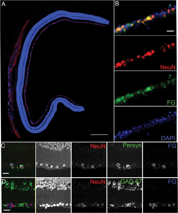
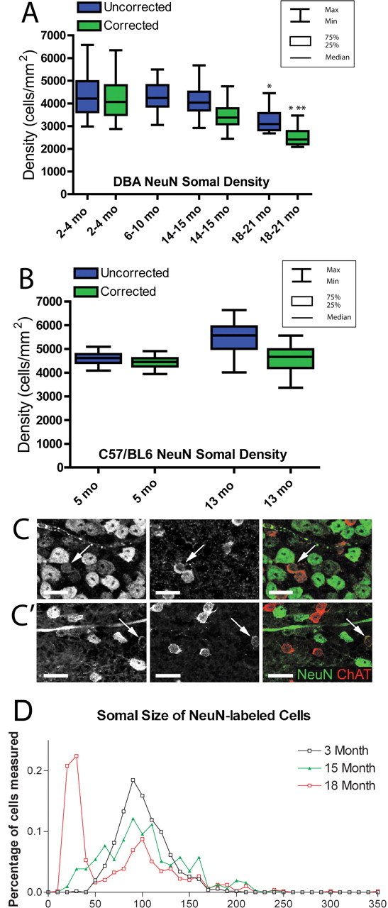
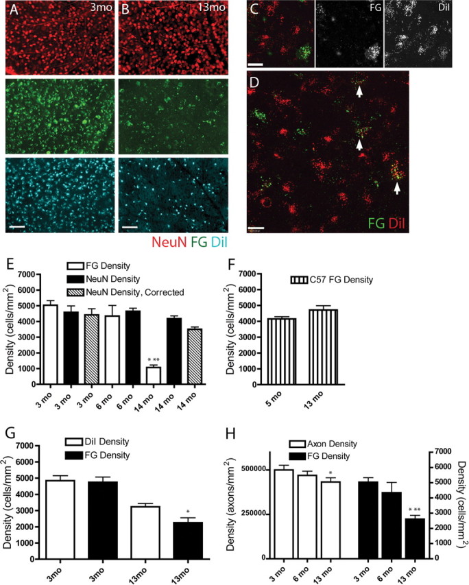
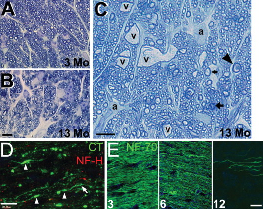
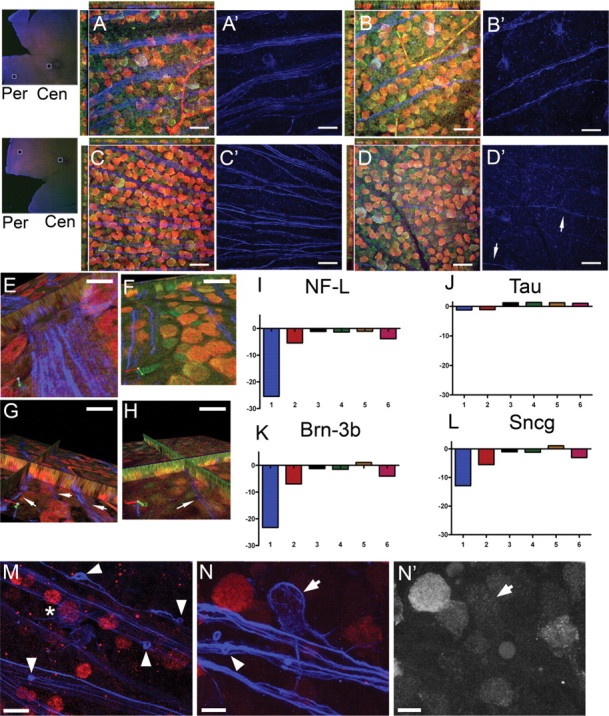
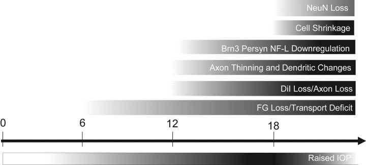
Glaucoma is characterized by retinal ganglion cell (RGC) pathology and a progressive loss of vision. Previous studies suggest RGC death is responsible for vision loss in glaucoma, yet evidence from other neurodegenerative diseases suggests axonal degeneration, in the absence of neuronal loss, can significantly affect neuronal function. To characterize RGC degeneration in the DBA/2 mouse model of glaucoma, we quantified RGCs in mice of various ages using neuronal-specific nuclear protein (NeuN) immunolabeling, retrograde labeling, and optic nerve axon counts. Surprisingly, the number of NeuN-labeled RGCs did not decline significantly until 18 months of age, at which time a significant decrease in RGC somal size was also observed. Axon dysfunction and degeneration occurred before loss of NeuN-positive RGCs, because significant declines in RGC number assayed by retrograde tracers and axon counts were observed at 13 months. To examine whether axonal dysfunction/degeneration affected gene expression in RGC axons or somas, NeuN and neurofilament-heavy (NF-H) immunolabeling was performed along with quantitative reverse transcription-PCR for RGC-specific genes in retinas of aged DBA/2 mice. Although these mice had similar numbers of NeuN-positive RGCs, the expression of neurofilament light, Brn-3b, and Sncg mRNA varied; this variation in RGC-specific gene expression was correlated with the appearance of NF-H immunoreactive RGC axons. Together, these data support a progression of RGC degeneration in this model of glaucoma, beginning with loss of retrograde label, where axon dysfunction and degeneration precede neuronal loss. This progression of degeneration suggests a need to examine the RGC axon as a locus of pathology in glaucoma.


Figures

Figure 1.
Figure 1.
Immunohistochemistry and in situ hybridization of RGCs in the DBA/2 retina showing that NeuN antibody predominantly labels RGCs. A, A vertical section through a 3-month-old DBA/2 retina immunolabeled with NeuN (red) and the nuclear stain DAPI (blue). Although occasionally observed in the inner nuclear layer, the majority of NeuN-positive cells are observed in the GCL. Scale bar, 250 μm. B, Higher-magnification view of double immunolabeling in the GCL for NeuN (red) and FG (green) with DAPI (blue) shows that all retrogradely labeled FluoroGold-positive RGCs are also NeuN positive. The panels broken out by fluorescence color channel allow one to visualize the variations in labeling intensity for both NeuN and FG. Scale bar, 50 μm. C, In situ hybridization combined with immunohistochemistry permitted colocalization of NeuN immunolabel, Sncg mRNA, and FG immunolabel in the GCL of a 3-month-old DBA/2 retina. All FG-positive RGCs were also NeuN positive and had Sncg mRNA. Breakout panels show the individual fluorescence color channels. Scale bar, 50 μm. D, In situ hybridization for the amacrine marker GAD-67 combined with immunohistochemistry for NeuN and FG show that all FG-positive cells are NeuN positive; FG-positive/NeuN-positive cells are GAD-67 negative. The individual fluorescence channels allow one to discern light NeuN immunolabeling in a small number of GAD-67 displaced amacrine cells. Scale bar, 50 μm.
Figure 2.
Figure 2.
Unbiased stereological cell quantification and somal size measurements to assess RGCs across the disease spectrum of the DBA/2. A, Box plot graph showing the density of NeuN-positive cells in the GCL both before (blue) and after (green) application of the NeuN/ChAT-positive amacrine cell correction factor. NeuN-positive densities (green) include only NeuN-positive RGCs. Boxes extend from the 25th to the 75th percentile, with a line at the median cell density. Error bars show the highest and lowest values. The 2–4 month group has the largest range of NeuN-positive cell density, whereas median density is similar across all age groups until 18–21 months. The oldest group has significantly lower NeuN-positive cell density than all age groups (*p < 0.01). There was no significant difference in corrected NeuN-positive cell density between the 2–4 and 14–15 month groups, and the 18–21 month group had significantly lower corrected NeuN-positive cell density than both of the other corrected age groups. This difference was greater between the 2–4 month group (**p < 0.001) than the 14–15 month group (*p < 0.01). A correction factor was not created for 6–10 month retinas, so this group is represented by total NeuN-positive cells only. B, Box plot graph illustrating NeuN-positive cells in the GCL of C57BL/6 control retinas at 5 and 13 months of age. NeuN-positive cell densities are represented as in A. There is no significant difference in corrected NeuN-positive RGC density (p > 0.1). C, C′, Immunolabeling for NeuN and ChAT in DBA/2 whole-mount retina at 3 (C) and 18 (C′) months illustrating a visible reduction in both number and size of NeuN-positive cells by 18 months. Arrows indicate amacrine cells with both NeuN and ChAT immunofluorescence. Scale bar, 20 μm. D, Histogram of NeuN-positive somal area as measured from retinal whole mounts at 3, 15, and 18 months. The 3 month retinas have nearly 20% NeuN-positive cells with a somal area ∼100 μm2; this percentage decreased with age. From 15–18 months, there is a significant increase in the number of NeuN-positive cells with somal areas <50 μm2 (p < 0.05).
Figure 3.
Figure 3.
RGC axon pathology assessed by comparing two retrograde labeling methods. A, Separate fields from 3 month (3mo) whole-mount retina immunolabeled with NeuN (red) and retrogradely labeled with FG (green) and DiI (blue). NeuN, FG, and DiI showed similar numbers across all retinas at 3 months. Scale bar, 100 μm. B, By 13 months (13mo), FG labeling (middle panel, green) in all retinas was dramatically reduced, and DiI labeling (bottom panel, blue) was reduced to a lesser extent, whereas NeuN labeling was unaffected (top panel, red). Immunohistochemistry for FG was done to improve the tracer signal; puncta smaller than somal size in the FG panel are nonspecific secondary antibodies within the tissue. Scale bar, 100 μm. C, High-magnification image of typical whole-mount retinal field of RGCs retrogradely labeled with FG (green) and DiI (red) at 14 months showing more DiI-positive than FG-positive cells. The cell in the lower right is FG and DiI positive. Scale bar, 20 μm. D, High-magnification field of DiI-FG retrogradely labeled DBA/2 retina whole-mount. Arrows point to RGCs with varying levels of FG and DiI labeling. The majority of cells are DiI positive. Scale bar, 20 μm. E, Graph of RGC density in DBA/2 retinal whole mounts as quantified with NeuN immunolabel and FG tracer. Data from 3 month (3 mo) and 14 month (14 mo) retinas are presented with (hash marked) and without (black) the displaced amacrine cell correction factor. No correction factor was available for 6 month (6 mo) DBA/2 data. NeuN and FG labeled a similar number of RGCs at 3 and 6 months, but at 14 months, FG labeling was markedly reduced compared with earlier time points (*) and compared with NeuN immunolabel in 14 month retinas (**) (p < 0.01). F, Graph of FG-labeled RGC density in retinal whole mounts of 5 month (5 mo) and 13 month (13 mo) C57BL/6 normal controls. Unlike DBA/2 retinas, there was no reduction in FG-labeled RGCs in older C57BL/6 controls. G, Graph of RGC density in DBA/2 retinal whole mounts labeled with both FG and DiI. FG and DiI labeling was similar at 3 months (3mo). By 13 months (13mo), there was a visible but not statistically significant decrease in DiI-positive RGCs, whereas the FG-positive (*) RGCs were significantly reduced (p < 0.05). H, Counts of RGC axons in the optic nerve from FG-quantified retinas showed a significant decrease by 13 months (*) (p < 0.05). FG retrograde labeling of RGCs at 13 months was significantly decreased relative to FG labeling at both 3 (*) and 6 (**) months (p < 0.05). As a percentage of the total, FG loss in the DBA/2 retinas was larger than axon loss in the corresponding optic nerves.
Figure 4.
Figure 4.
Optic nerves of DBA/2 mice show degenerative changes. A, Cross section of DBA/2 optic nerve at 3 months (3 Mo) processed for high-magnification light microscopy shows normal fascicle packing and regular myelin wraps on axons. B, Optic nerve cross section at 13 months (13 Mo) shows disruption of axon fascicles, increases in connective tissue septae, and lower axon numbers. C, Additional cross section of 13 month optic nerve showing vacuolization (v), large astrocytic processes (a), and arrows pointing to abnormal axon profiles. Arrowhead, Degenerating axon; wide arrow, myelin and axon pathology; narrow arrow, normal axon. Scale bars: (in B) A, B, 30 μm; C, 30 μm. D, Cholera toxin (CT) endocytosed by RGCs and transported through ganglion cell axons into the optic nerve showed numerous varicosities or swellings (arrowheads) and vacuolization (arrow) in addition to notable loss of NF-H immunoreactivity. Scale bar, 8 μm. E, Three, 6, and 12 month optic nerve immunolabeled with NF-70 showing accumulation of small neurofilaments from 3 to 6 months. By 12 months, NF-70 is nearly absent from many nerves. Scale bar, 10 μm.
Figure 5.
Figure 5.
Comparison of RGC-specific gene expression with NF-H immunolabel across 14 month DBA/2 retinas. A–D′, Photomicrographs taken from peripheral (Per) (3 month retina in A, A′; 14 month retina in C, C′) and central (Cen) (3 month retina in B, B′; 14 month retina in D, D′) whole-mount retina immunolabeled with NeuN (red) and NF-H (blue) and stained with NeuroTrace Nissl (green). A, B, This 3 month retina appeared to have a normal GCL, including healthy, well-labeled axon fascicles and strong NeuN immunolabeling. C, D, This 14 month retina did not exhibit loss of NeuN immunolabeling in the GCL, but there appeared to be a significant decrease in NF-H-positive axon fascicles. The axons present were thin and difficult to visualize (D′, arrows). E–H, A panel of photomicrographs from 14 month DBA/2 retinas immunolabeled with NeuN (red), NF-H (blue), and NeuroTrace Nissl (green). One half of the retina was removed for quantitative RT-PCR analysis of RGC-specific genes (I–L). E, F, The axon fascicles in this retina appeared robust, particularly near the central retina. The corresponding qPCR data for this retina are represented in lane 3 of I–L. G, H, This retina demonstrated a dramatic reduction in the size and number of axon fascicles (G, H, arrows) compared with the age- and IOP-matched retina in E and F. The corresponding qPCR data for this retina are represented in lane 1 of I–L. I–L, Quantitative PCR results for NF-L (I), tau (J), Brn3b (K), and Sncg (L) from six separate half retinas (lanes 1–6). There was no change in mRNA for tau, a neuronal protein, across these 14 month retinas, but there was significant downregulation of RGC-specific mRNAs for NF-L, Brn3b, and Sncg in some retinas, particularly the retina in lane 1. Photomicrographs of the retina in lane 3 (E, F) allow one to observe how retinas with normal RGCs and RGC axons had no changes in NF-L, tau, Brn3b, and Sncg mRNA (lane 3), whereas retinas with diminished axon fascicles in the nerve fiber layer (retina in G and H) also had RGCs with significantly downregulated RGC-specific genes (lane 1 in I–L). The magnitude of changes in NF-L, Brn3b, and Sncg are similar for individual retinas, suggesting a shared upstream regulation of these mRNAs. The variability of qPCR results illustrate the inconsistency in the glaucoma phenotype across 14 month DBA/2 mice. M–N′, Whole-mount retina immunolabeled with NeuN (red) and NF-H (blue) shows end bulbs of degenerating RGC axons (M, N, arrowheads) and downregulation of NeuN in RGCs. Prominent NF-H-positive RGCs were observed with moderate NeuN immunolabel (right of asterisk in M) or almost no NeuN immunolabel (arrow in N and N′). Scale bars: A–D′, 20 μm; E–H, M, 8 μm; N, N′, 5 μm.
Figure 6.
Figure 6.
Timeline of RGC changes in DBA/2 chronic secondary glaucoma. This timeline represents our conception of the pathogenesis of glaucoma from the point of view of the RGC. Gradients represent general onset and intensity of specified pathology over time. The timeline begins at 0 months; IOP increases commence at 6 months (Inman et al., 2006). The first observed decreases in FluoroGold labeling occur between 6 and 8 months in the DBA/2 and are significant at 13 months. Visible changes in DiI and significant decreases in axon number are evident by 13 months, suggesting that RGC axons may be dying back from target areas or degenerating at points between the superior colliculus and the retina 3–6 months after deficits in retrograde labeling are identified. Immunohistochemistry for NF-H shows axon thinning and degenerative end bulbs in retina and optic nerve at 12–14 months and beyond, whereas mRNA for RGC-specific genes such as Brn3b, Sncg, and NF-L decreases to a degree proportional to disease severity in this late period as well. Cell shrinkage has been observed at 15 months (Filippopoulos et al., 2006) and documented in this study at 18 months. The first significant decreases in NeuN-positive RGCs in this study occurred beginning at 18 months; this could represent downregulation of the NeuN protein or cell death.

References

    1. Anderson MG, Smith RS, Hawes NL, Zabaleta A, Chang B, Wiggs JL, John SW. Mutations in genes encoding melanosomal proteins cause pigmentary glaucoma in DBA/2J mice. Nat Genet. 2002;30:81–85. - PubMed
    1. Canola K, Angenieux B, Tekaya M, Quiambao A, Naash MI, Munier FL, Schorderet DF, Arsenijevic Y. Retinal stem cells transplanted into models of late stages of retinitis pigmentosa preferentially adopt a glial or a retinal ganglion cell fate. Invest Ophthalmol Vis Sci. 2007;48:446–454. - PMC - PubMed
    1. Chang B, Smith RS, Hawes NL, Anderson MG, Zabaleta A, Savinova O, Roderick TH, Heckenlively JR, Davisson MT, John SW. Interacting loci cause severe iris atrophy and glaucoma in DBA/2J mice. Nat Genet. 1999;21:405–409. - PubMed
    1. Chen YG, Zhang C, Chiang SK, Wu T, Tso MO. Increased nuclear factor-kappa B p65 immunoreactivity following retinal ischemia and reperfusion injury in mice. J Neurosci Res. 2003;72:125–131. - PubMed
    1. Coleman M. Axon degeneration mechanisms: commonality amid diversity. Nat Rev Neurosci. 2005;6:889–898. - PubMed

Publication types