Comparative Study
. 2008 Mar 12;28(11):2804-13.
doi: 10.1523/JNEUROSCI.5300-07.2008.

Adaptive control of saccades via internal feedback

Affiliations
Comparative Study

Adaptive control of saccades via internal feedback

Haiyin Chen-Harris et al. J Neurosci. .

Abstract

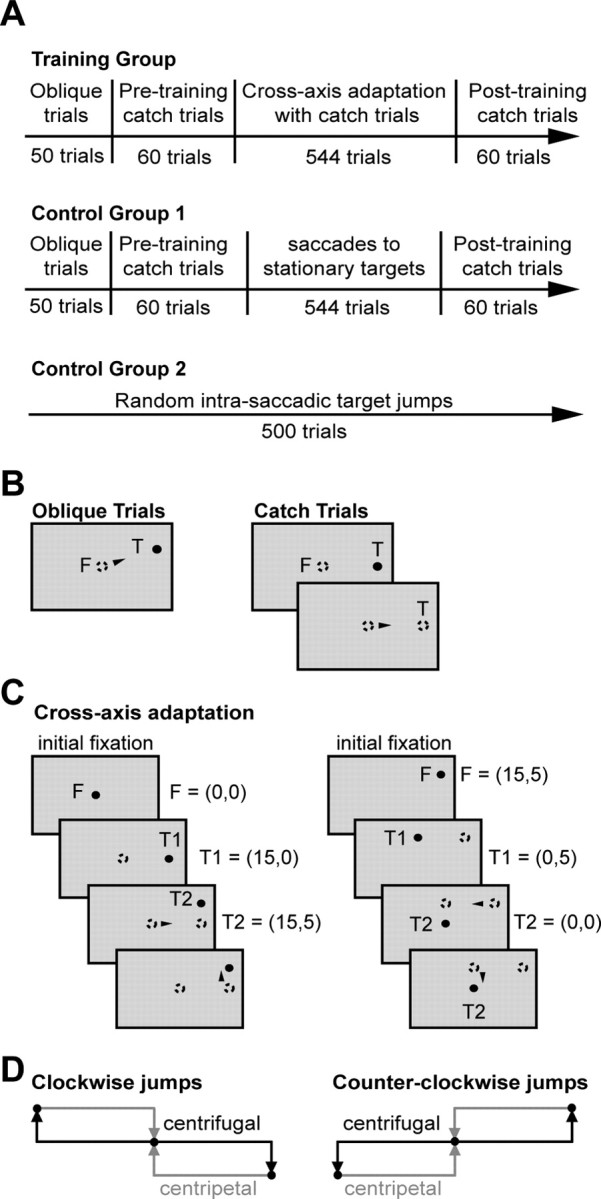
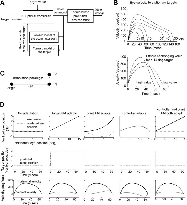
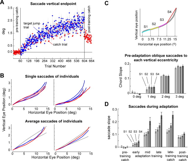
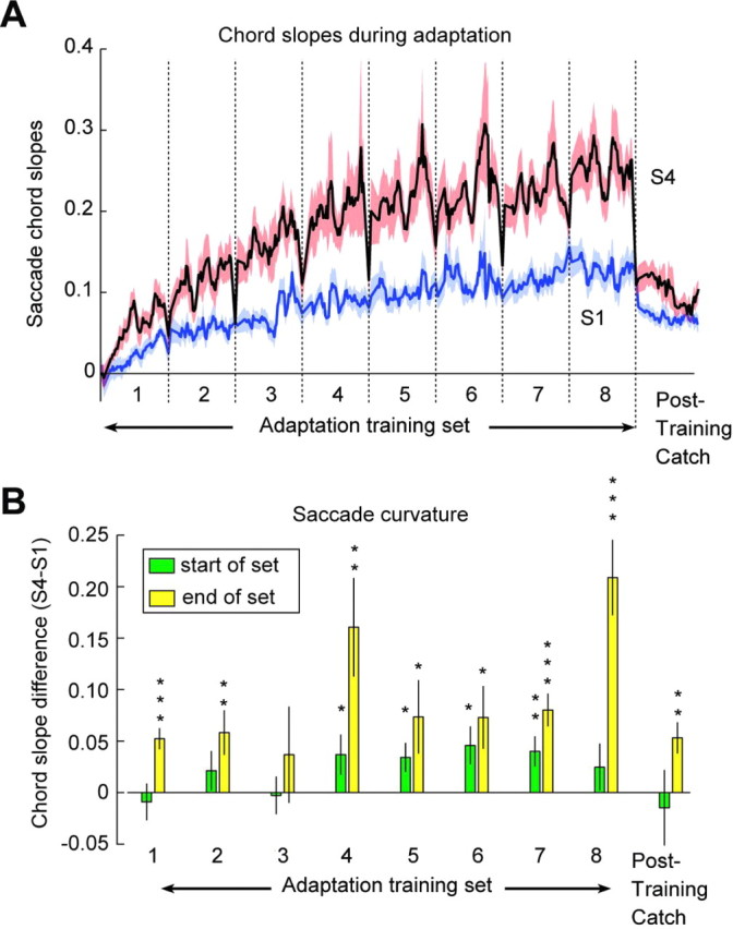
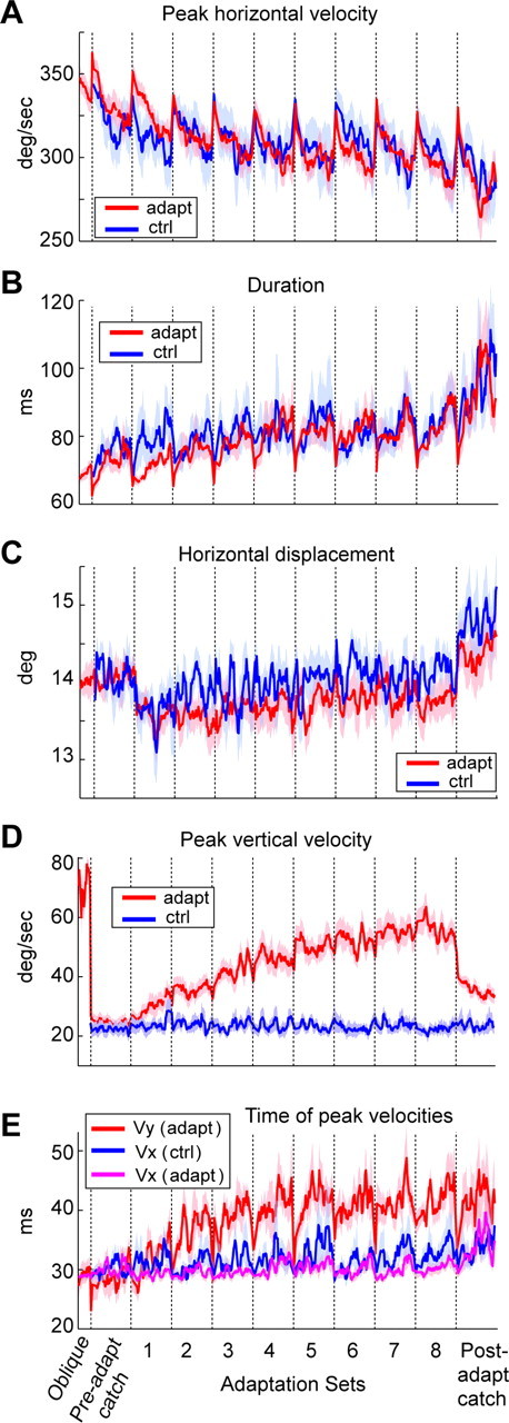
Ballistic movements like saccades require the brain to generate motor commands without the benefit of sensory feedback. Despite this, saccades are remarkably accurate. Theory suggests that this accuracy arises because the brain relies on an internal forward model that monitors the motor commands, predicts their sensory consequences, and corrects eye trajectory midflight. If control of saccades relies on a forward model, then the forward model should adapt whenever its predictions fail to match sensory feedback at the end of the movement. Using optimal feedback control theory, we predicted how this adaptation should alter saccade trajectories. We trained subjects on a paradigm in which the horizontal target jumped vertically during the saccade. With training, the final position of the saccade moved toward the second target. However, saccades became increasingly curved, i.e., suboptimal, as oculomotor commands were corrected on-line to steer the eye toward the second target. The adaptive response had two components: (1) the motor commands that initiated the saccades changed slowly, aiming the saccade closer to the jumped target. The adaptation of these earliest motor commands displayed little forgetting during the rest periods. (2) Late in saccade trajectory, another adaptive response steered it still closer to the jumped target, producing curvature. Adaptation of these late motor commands showed near-complete forgetting during the rest periods. The two components adapted at different timescales, with the late-acting component displaying much faster rates. It appears that in controlling saccades, the brain relies on an internal feedback that has the characteristics of a fast-adapting forward model.


Figures

Figure 1.
Figure 1.
Experimental paradigm. A, Chronology of the experiments. B, Target presentation sequence for oblique and catch trials. In oblique trials, targets were displayed randomly 15° lateral to the center LED and 0, 1, 2, 3, or 5° above (in the first quadrant) or below (in the third quadrant) the meridian. Each trial began with fixation (F) at the center LED (0°, 0°) for a random interval of 1–2 s, after which the center LED was extinguished and the target LED was turned on for 1 s. In catch trials, the target disappears at saccade onset. C, Target presentation sequence for a pair of adaptation trials during a counterclockwise adaptation experiment. Filled circles indicate currently illuminated LEDs, and open circles indicate previously illuminated LEDs. Arrowheads indicate when a saccade began. D, Target configurations for clockwise and counterclockwise cross-axis saccade adaptation experiments.
Figure 2.
Figure 2.
An optimal feedback control model of saccades. A, Schematic of the controller. B, Velocity profiles for typical saccades produced by the controller for various amplitudes. Each target has a position as well as a value. The top plot displays saccade velocities for a value α of 0.03 (Eq. 5). The bottom plot shows how the saccade velocity changes for a 15° target when the value of the target becomes smaller or larger (α = 0.0085, 0.015, and 0.03). C, Cross-axis saccade adaptation paradigm. While fixating the origin, target T1 is lit. As soon as the saccade begins, T1 is extinguished and T2 is lit. D, Comparisons of various implementations of cross-axis saccade adaptation. FM, Forward model. The teaching signal that guides adaptation is the retinal error at the end of the saccade. For the middle three columns, we assumed that endpoint errors are interpreted by the brain as inaccuracies in the control signals that generated the eye movements. This control signal depends on two structures: a controller and a forward model. If both structures adapt, then saccades will be straight to T2, despite the fact that the learner believes the eyes to be at T1. If the controller is the only structure that adapts, then saccades will show an initial vertical component, but then curve back toward T1 (because of the corrections imposed by the internal feedback from the FM). If the FM adapts but the controller does not, then the curvature will be toward T2. The curvature is again caused by the FM. Finally, it is possible that the brain interprets the errors as a jump in the position of the visual target (right-most column). In this case, the trajectories will be straight.
Figure 3.
Figure 3.
Adaptation in response to intrasaccadic target jump. A, Vertical endpoint of saccades during adaptation trials and postadaptation catch trials. Gray shading indicates SEM for each trial (n = 11 subjects). B, Saccades from two representative subjects. Top row, Primary saccade trajectories of the last three adaptation trials (blue) and the last two catch trials (red). Bottom row, Average primary saccade position in the last 10% of adaptation trials and the catch trials in the same trial range, overlaid on average trajectories to preadaptation control trials to oblique targets at (15°, 2°) (gray) and (15°, 3°) (gray). C, Top, Quantifying trajectory curvature. Each primary saccade trajectory was divided into four segments (four chords), and the slopes of the chords were labeled. Curvature is represented as relative change in slope from initial segment (white; S1) of the saccade to the final segment (black; S4). Bottom, Average preadaptation oblique saccades to each vertical eccentricity. Error bars represent SEM (n = 11). Before adaptation, oblique trials showed no tendency toward curvature (comparison of S2, S3, and S4 slopes with S1, p > 0.2 for all cases). D, During adaptation, S4 became increasingly larger than S1. S4 is significantly greater than S3 during the early, middle, and late 10% adaptation trials (48 trials each) at p = 0.002, p = 0.0003, and p = 0.000003 (1-tailed paired t test). “Late-training catch” refers to the 10 catch trials given during the last one-third of the adaptation block. S4 is significantly greater than S3 in the late-training and posttraining catch trials at p = 0.0002 and p = 0.002, respectively. Error bars represent SEM (n = 11).
Figure 4.
Figure 4.
The multiple timescales of adaptation. A, Moving average of the time course of S1 and S4 during adaptation (480 trials) and posttraining catch trials (60 trials). To highlight the rapid changes at the start of each set, we used a variable bin width (bw): bw = 2 trials for the first two trials in each set, bw = 4 for the next four trials, and bw = 6 for the rest of the set. Red and blue shaded areas represent SEM (n = 11). B, Curvature (chord slope S4 − S1) at the start and end of each set. The bin size is two saccades. Saccades that initiate each set have generally smaller curvature than saccades that complete the set. Eight of nine sets end with saccades that are curved (*p < 0.05; **p < 0.01; ***p < 0.001). Error bars represent SEM (n = 11).
Figure 5.
Figure 5.
Trial-dependent changes in saccade trajectories that were unrelated to adaptation. A, Saccade peak horizontal velocities. In both the control and the adaptation groups, the horizontal velocities showed a set structure suggesting a fatigue-like process in the horizontal motor commands that initiated the movements. B, Saccade durations. In both groups, saccade durations increased with a set structure consistent with a process that was highly correlated to the change in peak velocities (within-subject covariance between peak horizontal velocity and duration was significant at p < 10−5 for all subjects). C, Saccade horizontal amplitudes. Despite the reductions in peak horizontal velocity, horizontal amplitudes generally remained unchanged. D, Peak vertical velocity. In the adaptation group, endpoint errors induced changes in the vertical motor commands. These changes, as reflected by the peak vertical velocity, were specific to the adaptation group and did not exhibit the fatigue-like effects that we saw in the horizontal motor commands. Our noise level for saccade velocities is ∼22°/s. Therefore, the vertical component velocities shown in the control trials as well as the preadaptation catch trials reflect noise. Note the value for the control oblique saccades at the far left of the figure. E, Timing of the peak velocities in the horizontal (x) and vertical (y) directions for the adaptation experiment, and timing of peak horizontal velocity during the control experiment (timing of the peak vertical velocity was omitted because it was at noise levels). In the adaptation group, when horizontal velocity declined in each set, the adaptive response via internal feedback was an increase in the vertical motor command, resulting in a shift of the peak vertical velocity. Data in all panels are moving averages: fixed bin width (8) for oblique trial set and variable bin widths (Fig. 4A) for all other sets. Shades represent SEM.

Comment in

References

    1. Barash S, Melikyan A, Sivakov A, Zhang M, Glickstein M, Thier P. Saccadic dysmetria and adaptation after lesions of the cerebellar cortex. J Neurosci. 1999;19:10931–10939. - PMC - PubMed
    1. Boehnke SE, Berg DJ, Marino RA, Itti L, Munoz DP. Adaptation, habituation and dishabituation of visual responses in the superior colliculus. Soc Neurosci Abstr. 2007;33:617–12.
    1. Catz N, Dicke PW, Thier P. The compensation of saccadic fatigue is based on the adjustment of a Purkinje cell simple spike population. Soc Neurosci Abstr. 2006;32:345.22/S17.
    1. Deubel H. Adaptivity of gain and direction in oblique saccades. In: O'Regan JK, Levy-Schoen A, editors. Eye movements: from physiology to cognition. New York: Elsevier; 1987. pp. 181–190.
    1. Dinstein I, Hasson U, Rubin N, Heeger DJ. Brain areas selective for both observed and executed movements. J Neurophysiol. 2007;98:1415–1427. - PMC - PubMed

Publication types

LinkOut - more resources