Comparative Study
. 2008 Mar 12;28(11):2827-36.
doi: 10.1523/JNEUROSCI.4677-07.2008.

The mitochondrial protease AFG3L2 is essential for axonal development

Affiliations
Comparative Study

The mitochondrial protease AFG3L2 is essential for axonal development

Francesca Maltecca et al. J Neurosci. .

Abstract

The mitochondrial metalloprotease AFG3L2 assembles with the homologous protein paraplegin to form a supracomplex in charge of the essential protein quality control within mitochondria. Mutations of paraplegin cause a specific axonal degeneration of the upper motoneuron and, therefore, hereditary spastic paraplegia. Here we present two Afg3l2 murine models: a newly developed null and a spontaneous mutant that we found carrier of a missense mutation. Contrasting with the mild and late onset axonal degeneration of paraplegin-deficient mouse, Afg3l2 models display a marked impairment of axonal development with delayed myelination and poor axonal radial growth leading to lethality at P16. The increased severity of the Afg3l2 mutants is explained by two main molecular features that differentiate AFG3L2 from paraplegin: its higher neuronal expression and its versatile ability to support both hetero-oligomerization and homo-oligomerization. Our data assign to AFG3L2 a crucial role by linking mitochondrial metabolism and axonal development. Moreover, we propose AFG3L2 as an excellent candidate for motoneuron and cerebellar diseases with early onset unknown etiology.


Figures
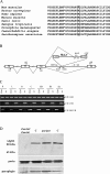
Figure 1.
Figure 1.
Molecular characterization of Afg3l2 mutant models. A, Partial sequence alignment of AAA domain of Afg3l2 orthologs. The mutant arginine residue (R389G, boxed) of Afg3l2 par/par mouse is highly conserved among different species. B, Schematic representation of provirus integration into intron 14 of the Afg3l2 gene. Arrows identify primers used (for details, see Materials and Methods). C, RT-PCR showing Afg3l2 Emv66/Emv66 and wild-type alleles expression in different tissues. K, Kidney; L, liver; H, heart; C, cerebellum; hB, hindbrain; fB, forebrain; Sp, spinal cord. D, Western blot analysis on isolated mitochondria from Afg3l2 Emv66/Emv66 (Emv66/Emv66), Afg3l2 par/par (par/par), and syngenic control mice (C). Anti-porin antibody is used to normalize sample loading.
Figure 2.
Figure 2.
Reduction of myelinated fibers in corticospinal tract. A, B, Semithin sections of spinal cord of Afg3l2 par/par mice and controls at P14. In the mutants the number of myelinated axons is dramatically reduced in the corticospinal tract. C, D, Ultrathin sections of corticospinal tract of Afg3l2 par/par mice and controls at P7. Myelinated axons are dramatically less represented in the mutants. E, F, At P12, ultrastructural analysis confirms the impressive reduction of myelinated fibers in the mutants compared with controls, where almost all axons are myelinated. Scale bar (in A): A, B, 20 μm; C, D, 1 μm; E, F, 2 μm. The graph shows counts of total myelinated axons in spinal cord from Afg3l2 par/par mice and controls at P14. A significant loss of axons in the mutant is visible. Comparing statistics are given as spinal region/t test p value: 1, corticospinal tract/0.014; 2, anterior funiculi/0.016; 3, anterolateral funiculi/0.008; 4, fasciculus gracilis/0.014. (supplemental text, available at www.jneurosci.org as supplemental material). Error bars represent ±SD; n = 3.
Figure 3.
Figure 3.
A–L, Extensive vacuolization of neuronal cell bodies. Semithin and ultrathin sections of DRGs (A–D), lumbar motoneurons (E–H), and cerebellum (I–L) of Afg3l2 par/par mice and controls at P14. Examples of multiple large intracytoplasmic vacuoles are indicated by empty polygons in the mutants (B, F, J). EM analysis shows giant mitochondria with swollen, disorganized cristae in the mutants (D, H, L, arrows) compared with controls (C, G, K). Scale bars: (in A) A, B, E, F, I, J, 20 μm; (in C) C, D, G, H, K, L, 1 μm.
Figure 4.
Figure 4.
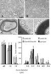
Sciatic nerve lacks large caliber axons. A, B, Semithin sections of sciatic nerve of control (A) and Afg3l2 par/par mouse (B) at P14. The reduced axonal diameter is evident. C, D, EM shows Schwann cell infoldings into the axon (C), and clustering of swollen mitochondria with poor residual cristae (D) is detected. E, At this age, compact myelin sheaths appear normal, with correct periodicity of major dense and intraperiod lines. Left graph shows counts of total, small (< 4 μm diameter), and large (> 4 μm diameter) myelinated axons in sciatic nerve from Afg3l2 par/par, Afg3l2 Emv66/Emv66, and control mice. A significant loss of large caliber axons appears in both mutants. Right graph shows averaged distribution of axon diameters from sciatic nerve of Afg3l2 par/par and Afg3l2 Emv66/Emv66 mice and controls. Axonal diameter distribution is skewed to 2–3 μm in the mutants. Statistical comparison with controls are as follows (class of axon diameter/t test p value): Afg3l2 Emv66/Emv66, <4 μm/0.005, >4 μm/0.021; Afg3l2 par/par, <4 μm/0.004, >4 μm/0.005. Error bars represent ±SD; n = 3. Scale bar (in A): A, B, 20 μm; C, 0.5 μm; D, E, 0.2 μm.
Figure 5.
Figure 5.
Delayed myelination is preceded by neuronal cell body vacuolization. A–H, Semithin sections of sciatic nerve at P5 (A, B), dorsal roots at P5 (C, D), ventral roots (E, F) at P1, and DRGs at P1 (G, H) of controls (left) and Afg3l2 par/par (right). Reduced number of myelinated fibers is evident in sciatic nerve (B) and dorsal and ventral roots (D and F, respectively). In the mutants, mitochondrial vacuolization is already present in DRGs at P1 (H). Scale bar, 20 μm.
Figure 6.
Figure 6.
A–C, Alteration of axoplasm structure. Sciatic nerve ultrathin preparations of control (A), Afg3l2 par/par mouse (B), and Afg3l2 Emv66/Emv66 (C) at P14. In the mutants, axoplasm appears poorly organized compared with the more even distribution observed in controls. Scale bar, 100 nm. Quantitative analysis (graph) reveals a reduction of neurofilament density in both mutants compared with controls. Afg3l2 par/par, p = 0.00001; Afg3l2 Emv66/Emv66 versus controls, p = 0.002 (t test). Error bars represent ±SD. An average of six axons in true cross sections were randomly selected for each genotype (n = 2).
Figure 7.
Figure 7.
Non-neuronal tissues are not significantly affected in Afg3l2 mutant mice. A–H, Cryostat (A–F) and ultrathin sections (G, H) from the quadriceps of Afg3l2 par/par mice and controls (A, D) at P10–P12. Hematoxylin/eosin staining (C) shows scattered atrophic fibers, an example of centrally placed nucleus, and some fibers with a basophilic cytoplasmic area. This area presents a red-purple stain with the modified trichrome staining (B), and intense reactivity with the SDH (E) and COX (F) staining. A quantitative evaluation of these findings in different muscles is represented in the graph, showing that aberrant fibers with either nuclei or mitochondria centrally positioned are very rare. Error bars represent ±SD; n = 3. Ultrastructural analysis of these sporadic altered fibers shows enlarged and morphologically disrupted mitochondria with distorted cristae, electron dense bodies, and lipid accumulation (G, H). Ultrathin sections of liver show no differences in mitochondrial morphology in mutant mice (J) compared with controls (I). Scale bars: (in A) A–F, 20 μm; (in G) G–J, 0.5 μm.
Figure 8.
Figure 8.
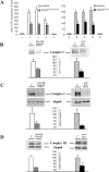
Respiratory chain complexes deficiencies in Afg3l2 mutant mice. A, ATP synthesis rates in mitochondria isolated from brain tissues. 1, No substrates (basal activity); 2, pyruvate and l-malate (complexes I, II, III, IV, and V); 3, glutamate and l-malate (complexes I, II, III, IV, and V); 4, rotenone and succinate (complexes II, III, IV, and V); 5, antimycin, ascorbate, and TMPD (complexes IV and V). Left graph, Afg3l2 Emv66/Emv66 substrate/t test p value: 2/0.008; 3/0.013; 4/0.044. Right graph, Afg3l2 par/par substrate/t test p value: 2/0.0004; 3/0.0002; 4/0.0006. B, BN-PAGE and in situ activity staining by nitroblue tetrazolium reduction assay. Comparison are relative to control; Afg3l2 Emv66/Emv66, p = 0.003; Afg3l2 par/par, p = 0.020 (t test). C, BN-PAGE-Immunoblot analysis of complex I revealed by anti-39 kDa antibody. Afg3l2 Emv66/Emv66, p = 0.024; Afg3l2 par/par, p = 0.0005 (t test). D, BN-PAGE-Immunoblot analysis of complex III revealed by anti-core2 (25.6 kDa subunit) antibody. Afg3l2 Emv66/Emv66, p = 0.011; Afg3l2 par/par, p = 0.004 (t test). Activity and protein amount of complex I and III were quantified by densitometric analysis; immunoblotting with Hsp60 antibody was used to verify equal loading. Error bars represent ±SD, n = 4.

References

    1. Atorino L, Silvestri L, Koppen M, Cassina L, Ballabio A, Marconi R, Langer T, Casari G. Loss of m-AAA protease in mitochondria causes complex I deficiency and increased sensitivity to oxidative stress in hereditary spastic paraplegia. J Cell Biol. 2003;163:777–787. - PMC - PubMed
    1. Blondet B, Duxson MJ, Harris AJ, Melki J, Guenet JL, Pincon-Raymond M, Rieger F. Nerve and muscle development in paralyse mutant mice. Dev Biol. 1989;132:153–166. - PubMed
    1. Bross P, Rugarli EI, Casari G, Langer T. Protein quality control in mitochondria and neurodegeneration in hereditary spastic paraplegia. Heidelberg: Springer; 2004.
    1. Casari G, De Fusco M, Ciarmatori S, Zeviani M, Mora M, Fernandez P, De Michele G, Filla A, Cocozza S, Marconi R, Dürr A, Fontaine B, Ballabio A. Spastic paraplegia and OXPHOS impairment caused by mutations in paraplegin, a nuclear-encoded mitochondrial metalloprotease. Cell. 1998;93:973–983. - PubMed
    1. Chang DT, Reynolds IJ. Mitochondrial trafficking and morphology in healthy and injured neurons. Prog Neurobiol. 2006;80:241–268. - PubMed

Publication types

Substances