. 2008 Jun;19(6):1106-15.
doi: 10.1681/ASN.2007070744. Epub 2008 Mar 12.

IL-6/IL-6R axis plays a critical role in acute kidney injury

Affiliations

IL-6/IL-6R axis plays a critical role in acute kidney injury

Yael Nechemia-Arbely et al. J Am Soc Nephrol. 2008 Jun.

Abstract

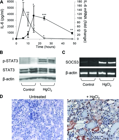
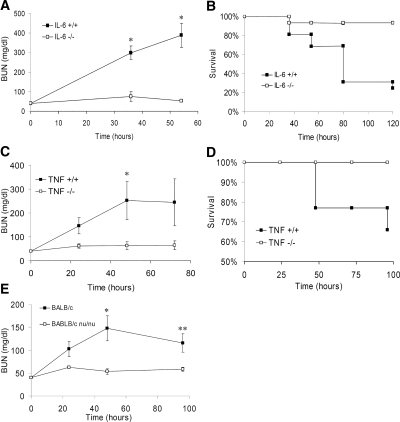
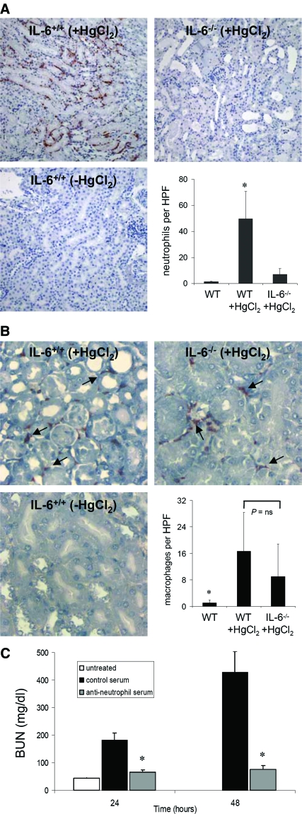
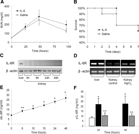
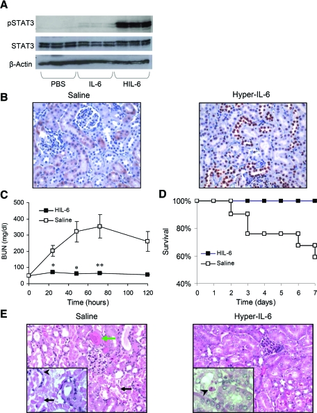
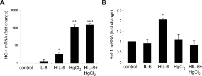
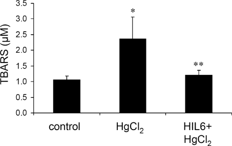
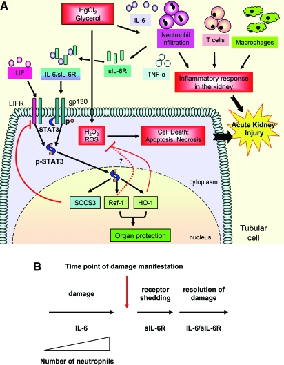
The response to tissue injury involves the coordination of inflammatory and repair processes. IL-6 expression correlates with the onset and severity of acute kidney injury (AKI), but its contribution to pathogenesis remains unclear. This study established a critical role for IL-6 in both the inflammatory response and the resolution of AKI. IL-6-deficient mice were resistant to HgCl2-induced AKI compared with wild-type mice. The accumulation of peritubular neutrophils was lower in IL-6-deficient mice than in wild-type mice, and neutrophil depletion before HgCl2 administration in wild-type mice significantly reduced AKI; these results demonstrate the critical role of IL-6 signaling in the injurious inflammatory process in AKI. Renal IL-6 expression and STAT3 activation in renal tubular epithelial cells significantly increased during the development of injury, suggesting active IL-6 signaling. Although a lack of renal IL-6 receptors (IL-6R) precludes the activation of classical signaling pathways, IL-6 can stimulate target cells together with a soluble form of the IL-6R (sIL-6R) in a process termed trans-signaling. During injury,serum sIL-6R levels increased three-fold, suggesting a possible role for IL-6 trans-signaling in AKI. Stimulation of IL-6 trans-signaling with an IL-6/sIL-6R fusion protein activated STAT3 in renal tubular epithelium and prevented AKI. IL-6/sIL-6R reduced lipid peroxidation after injury, suggesting that its protective effect may be largely mediated through amelioration of oxidative stress. In summary, IL-6 simultaneously promotes an injurious inflammatory response and, through a mechanism of trans-signaling, protects the kidney from further injury.


Figures

Figure 1.
Figure 1.
HgCl2 induces IL-6 expression and STAT3 activation. (A) IL-6 ELISA analysis of serum samples from mice treated with HgCl2 (6 mg/kg). Data are means ± SEM (n = 3 to 6 mice per time point). *P ≅ 0.004, **P ≅ 0.01 versus 0 h. Real-time PCR analysis of IL-6 mRNA in kidney taken from mice after treatment with HgCl2 (6 mg/kg; n = 3 to 6 mice per time point). *P < 0.05, **P ≅ 0.003, ***P ≅ 0.0001 versus 0 h. (B) Western blot analysis of p-STAT3 and STAT3 in the kidney 6 h after HgCl2-induced injury. (C) RT-PCR analysis of SOCS3 mRNA induction 6 h after HgCl2-induced injury. (D) p-STAT3 immunostaining in the kidney 24 h after HgCl2-induced injury reveals extensive STAT3 activation (red nuclear staining) in renal tubular epithelial cells. Magnification, ×200.
Figure 2.
Figure 2.
IL-6−/−, TNF-α−/−, and immune-deficient mice are partially resistant to HgCl2-induced AKI. (A) Renal function as indicated by BUN levels in IL-6+/+ and IL-6−/− mice administered HgCl2 (6 mg/kg). Data are means ± SEM of surviving mice. *P ≅ 0.001 versus IL-6+/+ mice (n = 14 to 15). (B) Kaplan-Meier survival plot of mice treated in A; log rank test, P ≅ 0.001 IL-6−/− versus IL-6+/+ mice. (C) Renal function in TNF-α+/+ (TNF+/+) and TNF-α−/− (TNF−/−) mice after HgCl2 administration. Data are means ± SEM of surviving mice. *P ≅ 0.04 (n = 5 to 9). (D) Kaplan-Meier survival plot of mice treated in C; log rank test, P ≅ 0.04 TNF+/+ versus TNF−/− mice. (E) Renal function as indicated by BUN levels in BALB/c and BALB/c nu/nu mice. Data are means ± SEM. *P ≅ 0.008, **P ≅ 0.03 (n = 8). No mortality was observed.
Figure 3.
Figure 3.
Neutrophilic infiltration to the renal parenchyma after injury is IL-6 dependent and promotes renal injury. (A) Few peritubular neutrophils are present at baseline in IL-6+/+ mice. Accumulation of neutrophils in peritubular capillaries at the inner cortex and outer medulla accompanied by neutrophilic extravasation into the renal interstitium is evident 24 h after HgCl2 administration. Peritubular accumulation of neutrophils in IL-6−/− mice after HgCl2 administration was significantly diminished compared with IL-6+/+ mice but not significantly different than in naïve IL-6+/+ mice. Quantification of renal neutrophilic infiltration in IL-6+/+ and IL-6−/− mice 24 h after HgCl2 administration is shown. Data are means ± SEM. *P ≅ 0.04 (n = 7) versus other groups. (B) Macrophage infiltration after AKI is not IL-6 dependent. Staining and quantification of renal macrophages in the inner cortex in IL-6+/+ and IL-6−/− mice 24 h after HgCl2 administration. Data are mean ± SEM. *P < 0.05 (n = 7) versus other groups. (C) Effect of neutrophil depletion on HgCl2-induced AKI. BUN levels in antineutrophil serum (□), control serum (▪), and untreated (baseline; □) are shown. Data are means ± SEM. *P ≅ 0.003 and ≅ 0.01 versus control serum + HgCl2–treated and untreated mice, respectively. HPF, high-power field. Magnification, ×200.
Figure 4.
Figure 4.
The absence of IL-6R expression in murine kidney precludes protection by IL-6 treatment, but sIL-6R is elevated after AKI. (A) Pretreatment with exogenous human IL-6 fails to protect mice from HgCl2-induced AKI. Renal function as indicated by BUN levels measured in mice treated with human IL-6 protein (20 μg, intravenously) or normal saline 4 h before administration of HgCl2 (6 mg/kg). Data are means ± SEM of surviving mice. P = NS, IL-6 treated versus control (n = 10). (B) Kaplan-Meier plot of survival in mice treated in A. Log rank test, P = NS. (C) Western blot analysis of IL-6R from normal murine liver and kidney and from kidney after HgCl2. The blots were stripped and reprobed for β-actin protein as a loading control. (D) IL-6R mRNA analysis by RT-PCR of RNA extracts from normal liver, normal kidney, and kidney tissue 6 h after HgCl2 administration. RT-PCR analysis of β-actin mRNA is shown for comparison. OD analysis: 141.7 ± 2.9 (liver), 28.8 ± 3.4 (normal kidney), and 58.7 ± 2.8 (kidney after HgCl2); P ≅ 0.008 for normal kidney versus kidney after HgCl2. (E) Serum sIL-6R ELISA analysis from HgCl2-treated (6 mg/kg) mice. Data are means ± SEM (n = 6 [48 h] to 12 mice [24 h]).*P < 0.00001, **P ≅ 0.002 versus 0 h. (F) Effect of neutrophil depletion on sIL-6R production after HgCl2-induced AKI. sIL-6R ELISA analysis on serum samples from antineutrophil serum (□) and control serum–treated mice (▪) after HgCl2 administration and from untreated mice (□) is shown. Data are means ± SD. *P ≅ 0.01 versus other groups; **P ≅ 0.01 versus control serum and P = NS versus untreated mice.
Figure 5.
Figure 5.
HIL-6 treatment protects mice from HgCl2-induced AKI. (A) Western blot analysis of p-STAT3 (Tyr 705) and STAT3 in the kidney after treatment with PBS, IL-6 (20 μg), or HIL-6 (4 μg) demonstrates the effect of HIL-6 on gp130 stimulation in the kidney. (B) pSTAT3 (Tyr705) immunostaining of kidney sections 1 h after HIL-6 treatment reveals strong STAT3 activation (expressed as nuclear staining), particularly in the distal tubules, medulla and glomeruli (strong staining), and proximal tubules (mild staining) (pSTAT3-positive tubular epithelial cells 307.7 ± 36.3 versus 0.0 ± 0.0 per HPF for HIL-6–and saline-treated mice, respectively; P ≅ 0.007). (C) Renal function in mice treated with HIL-6 or normal saline 4 h before administration of HgCl2. Data are means ± SEM. *P ≅ 0.002; **P ≅ 0.005 (n = 10). (D) Kaplan-Meier survival plot of mice treated as in B; log rank test, P ≅ 0.007, HIL-6 (n = 19) versus saline (n = 21). (E) Hematoxylin and eosin staining of paraffin-embedded renal tissue shows histologic changes 24 h after HgCl2 administration in mice pretreated with normal saline or HIL-6 (8 μg, intravenously) 4 h before HgCl2 administration. Saline-pretreated mice show extensive necrosis of tubular epithelial cells (black arrows), dilation of the tubular lumina filled with proteinaceous material (green arrow), and accumulation of neutrophils in the peritubular capillaries (arrowhead). HIL-6–pretreated mice display subtle tubular injury with occasional apoptotic tubular epithelial cells (arrowhead). Magnifications: ×400 in B and ×200 in E, inset; ×400 in E.
Figure 6.
Figure 6.
Effect of HIL-6 and IL-6 on the expression of oxidative stress response genes in normal and HgCl2-treated mice. (A) Real-time PCR analysis of HO-1 mRNA. *P ≅ 0.0006 versus control, **P ≅ 0.002; versus control; ***P ≅ 0.0001 versus control and P ≅ 0.03 versus HgCl2, (n = 4 to 7). (B) Real-time PCR analysis of Ref-1 mRNA. *P = 0.0002 versus other groups (n = 4 to 7).
Figure 7.
Figure 7.
HIL-6 prevents HgCl2-induced oxidative stress. Thiobarbituric acid reactive substances analysis of serum samples from mice 48 h after HgCl2-induced injury and mice treated with HIL-6 4 h before HgCl2 administration. Data are as means ± SD. *P ≅ 0.0002, HgCl2 (n = 13) versus control (n = 6); **P ≅ 0.0002, HIL-6+HgCl2 (n = 8) versus HgCl2.
Figure 8.
Figure 8.
Schematic view of the role of IL-6 and gp130 signaling in AKI. (A) IL-6 stimulates an immune-mediated inflammatory response involving neutrophil infiltration to the renal parenchyma that ultimately exacerbates renal injury. Stimulation of gp130 by IL-6 trans-signaling and leukemia inhibitory factor (LIF) or by therapeutic intervention with HIL-6 induces resistance to injury in tubular epithelial cells by activation of STAT3 and upregulation of redox-related genes including HO-1 and Ref-1. HO-1 is essential for protection against glycerol-induced but not HgCl2-induced AKI. These proteins may collaborate to prevent oxidative stress and reduce renal injury. ROS, reactive oxygen species. (B) A model for the dual role of gp-130 signaling in AKI. IL-6 promotes neutrophil infiltration via membrane-bound IL-6R and exacerbates renal injury. Neutrophils can release their membrane-bound IL-6R by shedding and promote protection by gp130 trans-signaling. This provides the basis of the dual role of IL-6 and IL-6/sIL-6R in tissue damage and protection.

References

    1. Brady HR, Brenner BM: Acute renal failure. In: Harrison's Principles of Internal Medicine, 16th Ed., edited by Kasper DL, Fauci AS, Longo DL, Braunwald E, Hauser SL, Jameson JL, New York, McGraw-Hill, 2005, pp 1644–1653
    1. Simmons EM, Himmelfarb J, Sezer MT, Chertow GM, Mehta RL, Paganini EP, Soroko S, Freedman S, Becker K, Spratt D, Shyr Y, Ikizler TA: Plasma cytokine levels predict mortality in patients with acute renal failure. Kidney Int 65: 1357–1365, 2004 - PubMed
    1. Vaidya VS, Shankar K, Lock EA, Dixon D, Mehendale HM: Molecular mechanisms of renal tissue repair in survival from acute renal tubule necrosis: Role of ERK1/2 pathway. Toxicol Pathol 31: 604–618, 2003 - PubMed
    1. Lemay S, Rabb H, Postler G, Singh AK: Prominent and sustained up-regulation of gp130-signaling cytokines and the chemokine MIP-2 in murine renal ischemia-reperfusion injury. Transplantation 69: 959–963, 2000 - PubMed
    1. Gadient RA, Patterson PH: Leukemia inhibitory factor, Interleukin 6, and other cytokines using the GP130 transducing receptor: Roles in inflammation and injury. Stem Cells 17: 127–137, 1999 - PubMed

Publication types