. 2008 Jun;19(6):1079-91.
doi: 10.1681/ASN.2007070737. Epub 2008 Mar 12.

Role of integrins in the assembly and function of hensin in intercalated cells

Affiliations

Role of integrins in the assembly and function of hensin in intercalated cells

Soundarapandian Vijayakumar et al. J Am Soc Nephrol. 2008 Jun.

Abstract

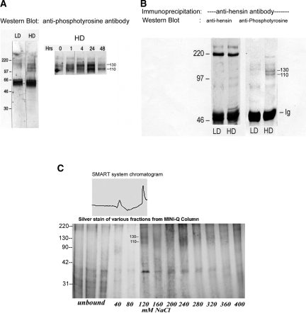
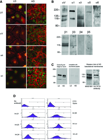
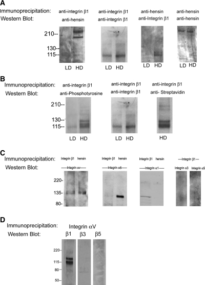
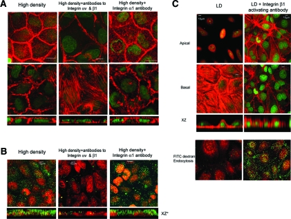
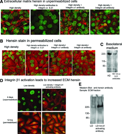
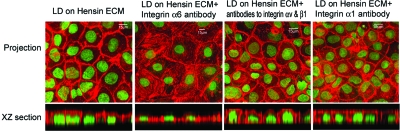
Epithelial differentiation proceeds in at least two steps: Conversion of a nonepithelial cell into an epithelial sheet followed by terminal differentiation into the mature epithelial phenotype. It was recently discovered that the extracellular matrix (ECM) protein hensin is able to convert a renal intercalated cell line from a flat, squamous shape into a cuboidal or columnar epithelium. Global knockout of hensin in mice results in embryonic lethality at the time that the first columnar cells appear. Here, antibodies that either activate or block integrin beta1 were used to demonstrate that activation of integrin alpha v beta 1 causes deposition of hensin in the ECM. Once hensin polymerizes and deposits into the ECM, it binds to integrin alpha 6 and mediates the conversion of epithelial cells to a cuboidal phenotype capable of apical endocytosis; therefore, multiple integrins play a role in the terminal differentiation of the intercalated cell: alpha v beta 1 generates polymerized hensin, and another set of integrins (containing alpha 6) mediates signals between hensin and the interior of the cells.


Figures

Figure 1.
Figure 1.
Identification of basolateral membrane proteins that interact with hensin. (A) Equal amounts of basolateral membrane proteins from low- (LD) and high-density (HD) cells were immunoblotted with anti-phosphotyrosine antibody (left). Two prominent protein bands at 110 and 130 kD were visible only in the HD phenotypes but not in the LD phenotypes (left). These two bands were also seen in HD cell lysates purified by wheat germ agglutinin and probed with anti-phosphotyrosine antibody, indicating these are glycosylated proteins (right). Maximal phosphorylation of these two bands was observed after 24 h of seeding (right). (B) Chemical cross-linking of the basolateral surface followed by immunoprecipitation with hensin antibodies. Only samples from HD cells showed 110- and 130-kD bands when probed with anti-phosphotyrosine antibody. The blots were stripped and reprobed with anti-hensin antibody showing equal amounts of hensin in both LD and HD samples (220-kD band). (C) Glycosylated membrane proteins from HD cells, purified using a Wheat Germ Lectin column, were adsorbed onto Mini-Q resin ion-exchange column and eluted with salt. The 110-/130-kD tyrosine-phosphorylated protein bands eluted at approximately 120 mM NaCl (corresponding to the second peak in the top panel). These bands were excised and analyzed using MALDI-TOF.
Figure 2.
Figure 2.
Integrin expression in clone C cells. (A) Expression patterns of integrin β1, αv, α6, and α1 in confluent monolayers of clone C cells seeded at LD and HD. Integrin staining is depicted in red, and nuclear staining with Sytox is shown in green. Integrin α3 staining was also observed by immunostaining but not depicted here. (B) Integrin expression in clone C cells was investigated by basolateral biotinylation followed by immunoprecipitation with integrin antibodies as indicated and Western blotted with anti-streptavidin antibodies (for αv and α3 in the top panel and all lanes in the bottom panel). In addition, Western blots were performed on biotin-streptavidin–purified basolateral membranes from HD cells with integrin α1, α5, and α6. (C) Western blots of cell lysates from LD and HD cells using the polyclonal antibody directed against extracellular domain of rabbit integrin β1 (left). (Right) Western blot using the same antibody in the presence of 600 ng/ml recombinant peptide immunogen. (D) Flow cytometry analysis of clone C cells with various integrin antibodies as indicated in the labels.
Figure 3.
Figure 3.
Integrin β1 binds to hensin and integrin αV and gets tyrosine phosphorylated in high density cells. (A) In the first panel, 140 μg of DTSSP–cross-linked cell protein from LD and HD phenotypes were immunoprecipitated with anti–integrin β1 CSAT antibodies, and the blots were probed with guinea pig anti-hensin antibody. In control experiment, membrane containing integrin β1 immunoprecipitates described in the first panel was stripped and reprobed with a different anti–integrin β1 antibody MAB1965 (second panel). Chemically cross-linked cell lysates from LD and HD cells were immunoprecipitated with goat anti-hensin/DMBT1 antibody and blotted with anti–integrin β1 MAB1965 antibody (third panel). Control experiment in which the same samples were probed with guinea pig anti-hensin antibody (fourth panel). (B) Tyrosine phosphorylation of integrin β1. (Left) Anti-phosphotyrosine Western blot of integrin β1 immunoprecipitates (described in A) from LD and HD. (Middle) Control Western blot with integrin β1 antibody. (Right) A streptavidin Western blot of biotinylated basolateral membrane proteins (from HD) that were immunoprecipitated with anti–integrin β1 antibody. (C) Interaction of integrin β1 with integrin α subunits was examined by Western blotting of integrin β1 immunoprecipitates with antibodies to integrin αv, α6, and α1 (left lane in first three panels) and α3 and α5 (last two lanes). Interaction of hensin with integrin α subunits was examined by Western blotting of hensin immunoprecipitates with antibodies to integrin αv, α6, and α1 (right lane in first three panels) from HD cells. (D) Interaction of integrin αv with integrin β subunits was examined by Western blotting of integrin αv immunoprecipitates with integrin β1 (first lane), integrin β3 (second lane), and integrin β5 (third lane) antibodies.
Figure 4.
Figure 4.
Role of integrin αv and β1 in terminal differentiation of clone C epithelia. (A) Clone C cells cultured at high density in media containing integrin αv and β1 function-blocking antibodies or integrin α1 function-blocking antibody. Apical and basal actin stained with phalloidin (red) and imaged by confocal microscopy showing en face and XZ optical sections. (B) HD cells cultured with or without integrin function-blocking antibodies as indicated in the labels were examined for their ability to endocytose fluorescein-dextran. Fluorescein-dextran in green and nuclear stain (Sytox red) in red. (C, top) Clone C cells seeded at low density and allowed to form confluent monolayers, when examined by F-Actin staining, have stress fibers on the basal surface and very little apical actin staining (left); however, when the same experiment is carried out in the presence of integrin β1–activating antibody P4G11, apical actin staining was observed with a marked difference in stress fiber staining on the basal surface. (Bottom) LD cells cultured in the absence or presence of integrin β1–activating antibody P4G11 were examined for their ability to endocytose green fluorescence (FITC) dextran. Nucleus was visualized with Sytox Red dye.
Figure 5.
Figure 5.
Integrins αv and β1 mediate ECM deposition of hensin. (A) ECM hensin staining: Clone C cells seeded at high density were cultured with various function-blocking integrin antibodies and exposed to anti-hensin antibodies (followed by rhodamine secondary antibodies) before fixation or permeabilization. Nuclear staining is shown in green. (B) Control experiment to examine intracellular hensin: Clone C cells seeded at high density were cultured with function-blocking integrin antibodies and exposed to anti-hensin antibodies after fixation and permeabilization. (C) Hensin secretion in basolateral medium: Equal amounts of combined basolateral medium from HD cells cultured in the presence and absence of integrin αv–and β1–blocking antibodies collected for 3 d was precleared with agarose beads, immunoprecipitated with anti-hensin (DMBT1) antibodies, and probed with anti-hensin (SRCR6/7) antibodies. (D) Clone C cells seeded at low density and cultured in the absence or presence of activating antibodies to integrin β1 and stained with hensin antibodies before (top) and after (bottom) permeabilization. (E) ECM was extracted from clone C cells seeded at identical low densities but cultured in the absence or presence of integrin β1–activating antibodies for 5 d. The ECM samples were then probed with Western blotting with guinea pig anti-hensin antibody.
Figure 6.
Figure 6.
Integrin α6 is a candidate for transmitting signals from ECM hensin to the cell interior. Effect of function-blocking α6 integrin antibodies on clone c cells seeded at low density on preformed hensin matrix and cultured for 5 d. Monolayers were then stained with Phalloidin (red) and Sytox (green), and the combined projection of all confocal sections is presented in these panels. XZ section represents a typical cross-section through the monolayer.

Comment in

References

    1. Vijayakumar S, Takito J, Gao X, Schwartz GJ, Al-Awqati Q: Differentiation of columnar epithelia: The hensin pathway. J Cell Science 119: 4797–4801, 2006 - PubMed
    1. Schwartz GJ, Barasch J, Al-Awqati Q: Plasticity of functional epithelial polarity. Nature 318: 368–371, 1985 - PubMed
    1. Edwards JC, van Adelsberg J, Rater M, Herzlinger D, Lebowitz J, Al-Awqati Q: Conditional immortalization of bicarbonate-secreting intercalated cells from rabbit. Am J Physiol 263: C521–C529, 1992 - PubMed
    1. van Adelsberg J, Edwards JC, Takito J, Kiss B, Al-Awqati Q: An induced extracellular matrix protein reverses the polarity of band 3 in intercalated epithelial cells. Cell 76: 1053–1061, 1994 - PubMed
    1. Vijayakumar S, Takito J, Hikita C, Al-Awqati Q: Hensin remodels the apical cytoskeleton and induces columnarization of intercalated epithelial cells: Processes that resemble terminal differentiation. J Cell Biol 144: 1057–1067, 1999 - PMC - PubMed

Publication types

LinkOut - more resources