. 2008 Mar 18;105(11):4312-7.
doi: 10.1073/pnas.0707370105. Epub 2008 Mar 12.

Anthrax lethal toxin and Salmonella elicit the common cell death pathway of caspase-1-dependent pyroptosis via distinct mechanisms

Affiliations

Anthrax lethal toxin and Salmonella elicit the common cell death pathway of caspase-1-dependent pyroptosis via distinct mechanisms

Susan L Fink et al. Proc Natl Acad Sci U S A. .

Abstract

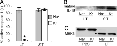
Caspase-1 cleaves the inactive IL-1beta and IL-18 precursors into active inflammatory cytokines. In Salmonella-infected macrophages, caspase-1 also mediates a pathway of proinflammatory programmed cell death termed "pyroptosis." We demonstrate active caspase-1 diffusely distributed in the cytoplasm and localized in discrete foci within macrophages responding to either Salmonella infection or intoxication by Bacillus anthracis lethal toxin (LT). Both stimuli triggered caspase-1-dependent lysis in macrophages and dendritic cells. Activation of caspase-1 by LT required binding, uptake, and endosome acidification to mediate translocation of lethal factor (LF) into the host cell cytosol. Catalytically active LF cleaved cytosolic substrates and activated caspase-1 by a mechanism involving proteasome activity and potassium efflux. LT activation of caspase-1 is known to require the inflammasome adapter Nalp1. In contrast, Salmonella infection activated caspase-1 through an independent pathway requiring the inflammasome adapter Ipaf. These distinct mechanisms of caspase-1 activation converged on a common pathway of caspase-1-dependent cell death featuring DNA cleavage, cytokine activation, and, ultimately, cell lysis resulting from the formation of membrane pores between 1.1 and 2.4 nm in diameter and pathological ion fluxes that can be blocked by glycine. These findings demonstrate that distinct activation pathways elicit the conserved cell death effector mechanism of caspase-1-mediated pyroptosis and support the notion that this pathway of proinflammatory programmed cell death is broadly relevant to cell death and inflammation invoked by diverse stimuli.


Conflict of interest statement

The authors declare no conflict of interest.

Figures

Fig. 1.
Fig. 1.
Lethal toxin and Salmonella stimulate caspase-1-dependent lysis of macrophages and DCs. (A–C) Macrophages were treated with PBS (A), S. typhimurium (S.T) (B), or LT (C), and active caspase-1 was identified by FAM-YVAD staining (green). Macrophages counterstained with TOPRO-3 (blue) were visualized by confocal fluorescence microscopy. (B and C) Discrete foci of active caspase-1 are indicated by filled arrowheads. (B) Bacteria stained by TOPRO-3 are indicated by open arrowheads. (D and E) Macrophages (MΦ) or DCs were treated with LT or S.T in the presence of the specific caspase-1 inhibitor YVAD or the negative control inhibitor zFA. LDH released by dying cells was quantified; means ± SD are shown. *, P < 0.05 versus medium.
Fig. 2.
Fig. 2.
LT requires acidification of intracellular compartments and catalytic activity to stimulate caspase-1. Macrophages were treated with PBS, LT, LT containing catalytically inactive E687C mutant LF, or S. typhimurium (S.T) in the presence or absence of 10 mM NH4Cl to inhibit the acidification of intracellular compartments. (A) Macrophages containing active caspase-1 were identified by FAM-YVAD staining; means ± SD are shown. *, P < 0.05 versus LT. (B) Mature IL-18 was detected in supernatants by Western blot.
Fig. 3.
Fig. 3.
Calcium is required for caspase-1 activation by LT: target cell interaction and LF translocation into the cytosol. Macrophages were treated with LT or S. typhimurium (S.T) in Ca2+-free medium with 1 mM EDTA or in medium containing 150 μM verapamil, a Ca2+ channel blocker. (A) Macrophages containing active caspase-1 were identified by FAM-YVAD staining; means ± SD are shown. *, P < 0.05 versus medium. (B) Cleaved LF substrate MEK3 (arrow) in LT-intoxicated macrophages was detected by Western blot. Where indicated, macrophages treated with detergent released membrane-enclosed LF and cleaved MEK3 was detected by Western blot in the lysates.
Fig. 4.
Fig. 4.
LT activation of caspase-1 requires proteasome activity subsequent to LF-mediated proteolytic events. (A) Macrophages treated with LT or S. typhimurium (S.T) in the presence of proteasome inhibitors (1 μM MG-132 or 5 μM lactacystin) were examined for active caspase-1 by FAM-YVAD staining; means ± SD are shown. *, P < 0.05 versus medium. (B) Mature IL-18 was detected in supernatants by Western blot. (C) Cleaved MEK3 (arrow) in LT intoxicated macrophages was detected as in Fig. 3.
Fig. 5.
Fig. 5.
Potassium efflux is required for caspase-1 activation by LT, but is dispensable for pyroptosis induced by Salmonella. Macrophages were treated with LT or S. typhimurium (S.T) in standard medium containing high Na+ or modified medium containing Na+ replaced by K+. (A) Macrophages containing active caspase-1 were identified by FAM-YVAD staining; means ± SD are shown. *, P < 0.05 versus Na+ medium. (B) Mature IL-18 was detected in supernatants by Western blot. (C) Cleaved MEK3 (arrow) in LT-intoxicated macrophages was detected as in Fig. 3.
Fig. 6.
Fig. 6.
Lethal toxin and Salmonella stimulate a common mechanism of lysis mediated by pore formation. (A–C) Macrophages were treated with LT, S. typhimurium (S.T), or 5 mM H2O2 in the presence of osmoprotectants of varying sizes (A and B) or glycine (B and C), which nonspecifically inhibits ion fluxes. (A and C) LDH released by dying cells was quantified; means ± SD are shown. *, P < 0.05 versus medium. (B) Macrophages containing active caspase-1 were identified by FAM-YVAD staining; means ± SD are shown.
Fig. 7.
Fig. 7.
Caspase-1-dependent DNA fragmentation occurs in LT-treated macrophages and during Salmonella infection. Macrophages were treated with PBS, LT, or S. typhimurium (S.T) in the presence of the specific caspase-1 inhibitor YVAD or the negative control inhibitor zFA. Macrophages containing damaged DNA were identified by TUNEL staining; means ± SD are shown. *, P < 0.05 versus medium.
Fig. 8.
Fig. 8.
Lethal toxin and Salmonella use distinct mechanisms to elicit the common pathway of caspase-1-dependent pyroptosis. (Upper) The LT complex consisting of PA and LF is taken up by macrophages in a Ca2+-dependent manner. Endosome acidification, which is blocked by NH4Cl, triggers a conformational change in PA, allowing translocation of LF into the cytosol. The Ca2+ channel blocker verapamil also inhibits LF translocation. In the cytosol, LF proteolytically cleaves MEK and other substrates, after which caspase-1 activation requires proteasome activity, potassium efflux, and the inflammasome protein Nalp1. Salmonella infection stimulates caspase-1 by an independent pathway requiring ligand(s) delivered by the bacterial type III secretion system (T3SS) and the inflammasome protein Ipaf. (Lower) Caspase-1 activated by both stimuli mediates a common pathway of pyroptosis: cell death featuring DNA fragmentation, secretion of activated inflammatory cytokines, and lytic release of inflammatory intracellular contents mediated by the formation of membrane pores between 1.1 and 2.4 nm in diameter. Osmotic lysis during pyroptosis is blocked by osmoprotectants and the cytoprotective agent glycine.

References

    1. Li P, et al. Mice deficient in IL-1 beta-converting enzyme are defective in production of mature IL-1 beta and resistant to endotoxic shock. Cell. 1995;80:401–411. - PubMed
    1. Wang W, et al. Endotoxemic acute renal failure is attenuated in caspase-1-deficient mice. Am J Physiol. 2005;288:F997–F1004. - PubMed
    1. Frantz S, et al. Targeted deletion of caspase-1 reduces early mortality and left ventricular dilatation following myocardial infarction. J Mol Cell Cardiol. 2003;35:685–694. - PubMed
    1. Melnikov VY, et al. Impaired IL-18 processing protects caspase-1-deficient mice from ischemic acute renal failure. J Clin Invest. 2001;107:1145–1152. - PMC - PubMed
    1. Siegmund B, Lehr HA, Fantuzzi G, Dinarello CA. IL-1 beta-converting enzyme (caspase-1) in intestinal inflammation. Proc Natl Acad Sci USA. 2001;98:13249–13254. - PMC - PubMed

Publication types

LinkOut - more resources