. 2008 Mar 18;105(11):4247-52.
doi: 10.1073/pnas.0800276105. Epub 2008 Mar 12.

Distinct effects of the recurrent Mlh1G67R mutation on MMR functions, cancer, and meiosis

Affiliations

Distinct effects of the recurrent Mlh1G67R mutation on MMR functions, cancer, and meiosis

Elena Avdievich et al. Proc Natl Acad Sci U S A. .

Abstract

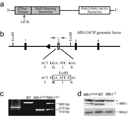
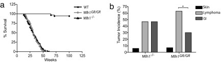
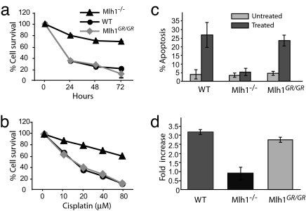
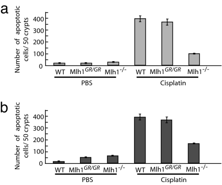
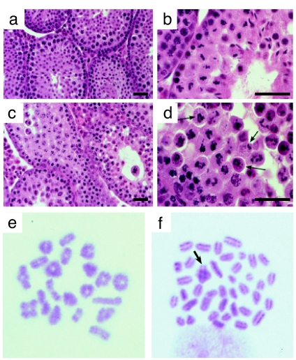
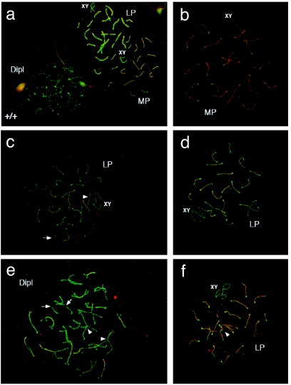
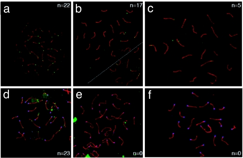
Mutations in the human DNA mismatch repair (MMR) gene MLH1 are associated with hereditary nonpolyposis colorectal cancer (Lynch syndrome, HNPCC) and a significant proportion of sporadic colorectal cancer. The inactivation of MLH1 results in the accumulation of somatic mutations in the genome of tumor cells and resistance to the genotoxic effects of a variety of DNA damaging agents. To study the effect of MLH1 missense mutations on cancer susceptibility, we generated a mouse line carrying the recurrent Mlh1(G67R) mutation that is located in one of the ATP-binding domains of Mlh1. Although the Mlh1(G67R) mutation resulted in DNA repair deficiency in homozygous mutant mice, it did not affect the MMR-mediated cellular response to DNA damage, including the apoptotic response of epithelial cells in the intestinal mucosa to cisplatin, which was defective in Mlh1(-/-) mice but remained normal in Mlh1(G67R/G67R) mice. Similar to Mlh1(-/-) mice, Mlh1(G67R/G67R) mutant mice displayed a strong cancer predisposition phenotype. However, in contrast to Mlh1(-/-) mice, Mlh1(G67R/G67R) mutant mice developed significantly fewer intestinal tumors, indicating that Mlh1 missense mutations can affect MMR tumor suppressor functions in a tissue-specific manner. In addition, Mlh1(G67R/G67R) mice were sterile because of the inability of the mutant Mlh1(G67R) protein to interact with meiotic chromosomes at pachynema, demonstrating that the ATPase activity of Mlh1 is essential for fertility in mammals.


Conflict of interest statement

The authors declare no conflict of interest.

Figures

Fig. 1.
Fig. 1.
The generation of Mlh1G67R mutant mice. (a) Domain structure of Mlh1. The location of the G67R mutation is indicated. (b) Schematic representation of the modified Mlh1G67R genomic locus and sequence characteristics of the G67R mutation. (c) PCR genotyping of tail DNA from Mlh1G67R mutant mice. Mlh1G67R/+, heterozygous mice; Mlh1G67R/G67R, homozygous mutant mice. After EcoRI restriction digestion of the PCR product, the WT allele is indicated by an 880-bp fragment and the Mlh1G67R mutant allele by 470- and 410-bp restriction fragments. (d) Western blot analyses of thymocyte cell extracts using anti-Mlh1 and anti-Msh2 antibodies.*, Unspecific protein recognized by the Mlh1 antisera.
Fig. 2.
Fig. 2.
Survival and tumor incidence in Mlh1 mutant mice. (a) Survival of Mlh1 mutant mice. Significantly reduced survival of Mlh1−/− and Mlh1G67R/G76R mice compared with WT mice (P < 0.0001; log-rank test) is shown. There was no difference in survival between Mlh1−/− and Mlh1G67R/G76R mice (P = 0.915; log-rank test). (b) Tumor incidence in Mlh1−/− and Mlh1G67R/G67R mutant mice. Reduced gastrointestinal (GI) tumor incidence in Mlh1G67R/G67R mice (P < 0.0296) is shown.
Fig. 3.
Fig. 3.
Cisplatin sensitivity and apoptosis in Mlh1G67R/G67R MEF cells. MEF strains of the various Mlh1 genotypes were exposed to cisplatin for different time periods or at varying concentrations. (a) Survival of cells after exposure with 40 μM cisplatin at different time intervals. (b) Survival of cells after 48-h exposure at different cisplatin concentrations. (c) Apoptotic response to cisplatin treatment (20 μM cisplatin for 24 h) measured by TUNEL. (d) G2/M cell-cycle arrest in Mlh1G67R/G67R MEF cells. The percentage of cells in G0/G1 and G2/M were calculated for three different MEF strains for indicated genotypes. The results are shown as the fold increase of G2/M cells in treated over untreated MEFs for each genotype.
Fig. 4.
Fig. 4.
Cisplatin-induced apoptotic response of epithelial cells in the intestinal mucosa in Mlh1 mutant mice. TUNEL-positive cells 24 h after cisplatin exposure (10 mg/kg of body weight). (a) Small intestine. (b) Large intestine.
Fig. 5.
Fig. 5.
Analysis of Mlh1G67R/G67R mutant testes reveals spermatogenic failure at or before metaphase of the first meiotic division. (a–d) H&E staining of testis sections from WT (a and b) and Mlh1G67R/G67R (c and d) males, showing normal progression of spermatogenesis in WT and failure to progress beyond metaphase I in mutant adult testes. Aberrant spindle configurations are observed in testis sections from males (arrows). (Scale bar, 100 μm.) (e and f) Metaphase spreads from WT (e) and Mlh1G67R/G67R (f) spermatocytes show abnormal metaphase configurations in the mutant mice. Almost all chromosomes are univalent, with only very few crossovers remaining (arrow) in Mlh1G67R/G67R spermatocytes.
Fig. 6.
Fig. 6.
Premature chromosome desynapsis in Mlh1G67R/G67R mice. Sycp1 (red) and Sycp3 (green) staining shows pachytene to diplotene chromosome configurations in WT (a) and Mlh1G67R/G67R (b–f) mouse spermatocytes. Mutant animals show premature separation of some chromosomes, particularly the XY in midpachynema (MP) and longer chromosomes in late pachynema (LP) to early diplonema (arrowheads in c) with abnormal intrahomolog associations being evident at late pachynema and diplonema (Dipl) (arrowheads in e and f). Broken synaptonemal complexes are also common (arrows in c and e). (See also larger images in SI Fig. 12 in SI Appendix).
Fig. 7.
Fig. 7.
Localization of Mlh1 (a–c) and Mlh3 (d–f) on meiotic chromosomes. (a–c) Localization of Mlh1 (green foci) on pachytene chromosome spreads from WT (a) and Mlh1G67R/G67R (b and c) males reveals a dramatic decrease in frequency and intensity of Mlh1 staining in the Mlh1G67R/G67R mutant animals. Many cells show a complete absence of Mlh1G67R staining (data not shown), but others show distinct Mlh1G67R reduction as exemplified in b and c. (d–f) Mlh3 staining (green foci) of WT (d) and Mlh1G67R/G67R (e and f) males reveals a complete absence of Mlh3 staining in pachytene spreads from Mlh1G67R/G67R mutant animals. (See also larger images in SI Fig. 13 in SI Appendix). Synaptonemal complexes are stained with anti Sycp3 antiserum (red), and centromeres are detected by CREST autoimmune serum (blue).

References

    1. Peltomaki P, Vasen HF. Mutations predisposing to hereditary nonpolyposis colorectal cancer: Database and results of a collaborative study. The International Collaborative Group on Hereditary Nonpolyposis Colorectal Cancer. Gastroenterology. 1997;113:1146–1158. - PubMed
    1. Yan H, et al. Conversion of diploidy to haploidy. Nature. 2000;403:723–724. - PubMed
    1. Kunkel TA, Erie DA. DNA mismatch repair. Annu Rev Biochem. 2005;74:681–710. - PubMed
    1. Schofield MJ, Hsieh P. DNA mismatch repair: Molecular mechanisms and biological function. Annu Rev Microbiol. 2003;57:579–608. - PubMed
    1. Kolas NK, Cohen PE. Novel and diverse functions of the DNA mismatch repair family in mammalian meiosis and recombination. Cytogenet Genome Res. 2004;107:216–231. - PubMed

Publication types

MeSH terms

LinkOut - more resources