. 2008 Mar 18;105(11):4271-6.
doi: 10.1073/pnas.0711875105. Epub 2008 Mar 12.

PPARdelta regulates multiple proinflammatory pathways to suppress atherosclerosis

Affiliations

PPARdelta regulates multiple proinflammatory pathways to suppress atherosclerosis

Grant D Barish et al. Proc Natl Acad Sci U S A. .

Abstract

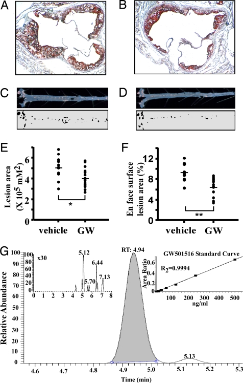
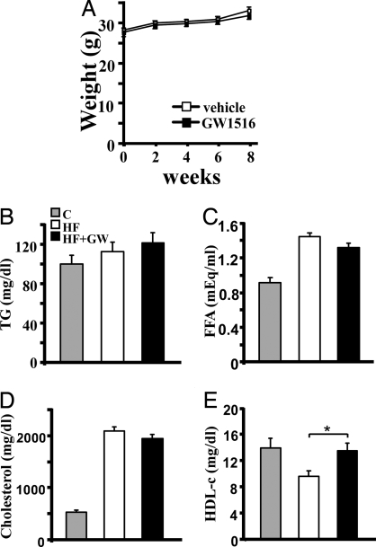
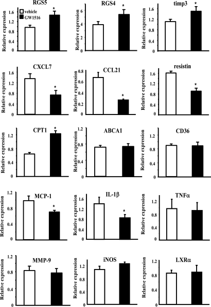
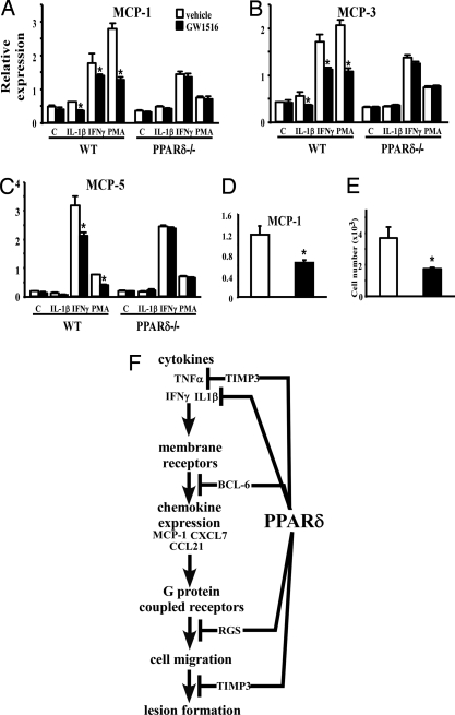
Lipid homeostasis and inflammation are key determinants in atherogenesis, exemplified by the requirement of lipid-laden, foam cell macrophages for atherosclerotic lesion formation. Although the nuclear receptor PPARdelta has been implicated in both systemic lipid metabolism and macrophage inflammation, its role as a therapeutic target in vascular disease is unclear. We show here that orally active PPARdelta agonists significantly reduce atherosclerosis in apoE(-/-) mice. Metabolic and gene expression studies reveal that PPARdelta attenuates lesion progression through its HDL-raising effect and anti-inflammatory activity within the vessel wall, where it suppresses chemoattractant signaling by down-regulation of chemokines. Activation of PPARdelta also induces the expression of regulator of G protein signaling (RGS) genes, which are implicated in blocking the signal transduction of chemokine receptors. Consistent with this, PPARdelta ligands repress monocyte transmigration and macrophage inflammatory responses elicited by atherogenic cytokines. These results reveal that PPARdelta antagonizes multiple proinflammatory pathways and suggest PPARdelta-selective drugs as candidate therapeutics for atherosclerosis.


Conflict of interest statement

The authors declare no conflict of interest.

Figures

Fig. 1.
Fig. 1.
PPARδ activation protects against atherosclerosis. (A and B) Representative oil red O-stained sections of aortic valves from vehicle (A) or GW501516 (GW)-treated (B) apoE−/− mice. Ten-week-old male apoE−/− mice on an atherogenic diet were gavaged daily with either vehicle or GW, a high-affinity PPARδ agonist, at 2 mg·kg−1·day−1 for 8 weeks. (C and D) En face lesion area in representative vehicle (C) or GW-treated (D) aortas. (E and F) Quantitative analysis of the lesion size in aortic valves (E) and aortas (F) (n = 15). GW treatment significantly reduced lesion sizes. *, P < 0.05; **, P < 0.005 Mann–Whitney U test. (G) LC-MS/MS quantitation of GW drug levels in pooled serum from vehicle (Inset Left) versus drug-treated mice. Standard curve for quantitation of GW levels (Inset Right).
Fig. 2.
Fig. 2.
PPARδ increases HDL-c in apoE−/− mice. (A) GW does not affect weight gain. (B–D) Similar levels of serum triglyceride, free fatty acid (FFA), and cholesterol are observed between vehicle- and GW-treated groups. C, chow diet; HF, high-fat diet; HF+GW, high-fat diet with GW treatment. (E) An increase in HDL cholesterol occurs in GW-treated apoE−/− mice. *, P < 0.05.
Fig. 3.
Fig. 3.
Real-time PCR analyses of potential PPARδ target genes in the aorta. Verification of PPARδ regulated genes identified by DNA array experiments and examination of additional atherosclerosis-related genes by using whole aorta RNA from vehicle- or GW-treated apoE−/− mice. Results are presented as mean (of three mice) ± SEM. *, P < 0.05.
Fig. 4.
Fig. 4.
Activation of PPARδ attenuates chemoattractant signaling. (A–C) Real-time PCR demonstrating ligand treatment suppresses chemokine expression induced by IL-1β, IFNγ, and PMA in a receptor-dependent manner. Thioglycollate-elicited peritoneal macrophages were isolated from wild-type (wt) or PPARδ−/− mice and stimulated with the indicated stimulant in the presence or absence of ligand. (D) Reduced MCP-1 expression in peritoneal macrophages isolated from ligand treated apoE−/− mice. (E) PPARδ ligand treatment inhibits monocyte transmigration. THP-1 monocytes pretreated with vehicle or GW were examined for their ability to migrate through an endothelial cell monolayer along an MCP-1 gradient. (F) Schematic demonstrating that PPARδ inhibits inflammatory signaling through multiple downstream effectors. We have previously shown that PPARδ regulates the expression of the cytokine IL-1β and ligand-bound PPARδ releases the transcriptional repressor BCL-6 to repress the expression of chemokines such as MCP-1. In this study, RGSs and TIMP-3 are shown to be up-regulated by PPARδ activation. RGSs block chemokine receptor signal transduction, whereas TIMP-3 inhibits TNFα shedding and cell migration. *, P < 0.05.

References

    1. Glass CK, Witztum JL. Atherosclerosis. The road ahead. Cell. 2001;104:503–516. - PubMed
    1. Libby P, Ridker PM, Maseri A. Inflammation and atherosclerosis. Circulation. 2002;105:1135–1143. - PubMed
    1. Gosling J, et al. MCP-1 deficiency reduces susceptibility to atherosclerosis in mice that overexpress human apolipoprotein B. J Clin Invest. 1999;103:773–778. - PMC - PubMed
    1. Gu L, et al. Absence of monocyte chemoattractant protein-1 reduces atherosclerosis in low density lipoprotein receptor-deficient mice. Mol Cell. 1998;2:275–281. - PubMed
    1. Dawson TC, Kuziel WA, Osahar TA, Maeda N. Absence of CC chemokine receptor-2 reduces atherosclerosis in apolipoprotein E-deficient mice. Atherosclerosis. 1999;143:205–211. - PubMed

Publication types

MeSH terms