. 2008 May 15;111(10):4997-5007.
doi: 10.1182/blood-2007-08-108597. Epub 2008 Mar 12.

TNF primes endothelial cells for angiogenic sprouting by inducing a tip cell phenotype

Affiliations

TNF primes endothelial cells for angiogenic sprouting by inducing a tip cell phenotype

Richard C A Sainson et al. Blood. .

Abstract

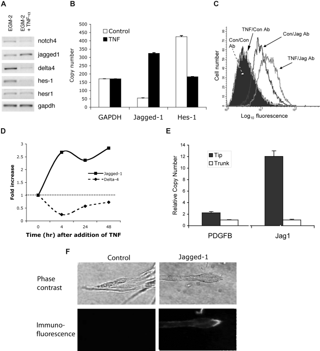
Pathological angiogenesis associated with wound healing often occurs subsequent to an inflammatory response that includes the secretion of cytokines such as tumor necrosis factor (TNF). Controversy exists on the angiogenic actions of TNF, with it being generally proangiogenic in vivo, but antiangiogenic in vitro. We find that whereas continuous administration of TNF in vitro or in vivo inhibits angiogenic sprouting, a 2- to 3-day pulse stimulates angiogenesis by inducing an endothelial "tip cell" phenotype. TNF induces the known tip cell genes platelet-derived growth factor B (PDGFB) and vascular endothelial cell growth factor receptor-2 (VEGFR2), while at the same time blocking signaling through VEGFR2, thus delaying the VEGF-driven angiogenic response. Notch signaling regulates tip cell function, and we find that TNF also induces the notch ligand jagged-1, through an NFkappaB-dependent mechanism. Enrichment of jagged-1 in tip cells was confirmed by immunofluorescent staining as well as by laser capture microdissection/quantitative reverse-transcription-polymerase chain reaction (qRT-PCR) of tip cells sprouting in vitro. Thus, in angiogenesis, the temporal expression of TNF is critical: it delays angiogenesis initially by blocking signaling through VEGFR2, but in addition by inducing a tip cell phenotype through an NFkappaB-dependent pathway, it concomitantly primes endothelial cells (ECs) for sprouting once the initial inflammatory wave has passed.


Figures

Figure 1
Figure 1
A pulse of TNF promotes angiogenesis in vitro. (A) EC-coated beads in fibrin gels were treated with TNF (10 ng/mL) or PBS control and photographed after 4 days on an Olympus IX70 inverted phase-contrast microscope using a 4×/0.13 NA objective and an Optronics digital camera. High-resolution images were magnified in Photoshop. Control cultures showed robust sprouting, whereas TNF almost completely blocked sprouting. One of multiple similar experiments. (B) Cultures were established as in panel A and TNF was added at day 0, 2, or 4. The number of sprouts was counted at day 6. Shown are means and SD; *P < .005 relative to control, by Student t test. One of 2 similar experiments. (C) Cultures were established as above and treated for 2 days with or without TNF (10 ng/mL). TNF-containing medium was then removed and fresh medium lacking TNF was added every 2 days. Sprouts were counted at the indicated times. Shown are means and SD; *P < .005 relative to control, by Student t test. One of 3 similar experiments. (D) ECs (4 × 10⁴) were plated in 24-well plates in the presence or absence of TNF (10 ng/mL) and counted at the indicated times. *P < .005 relative to control, by Student t test. One of 2 similar experiments. (E) TNF blocks VEGF signaling acutely. ECs were pretreated with TNF (10 ng/mL) for 2 days, rested for 1 day, and then given VEGF (10 ng/mL) for 4 hours (TNF then VEGF), or were given TNF for 3 days and then TNF + VEGF for the last 4 hours (TNF + VEGF), or were given VEGF alone for 4 hours (VEGF) or left untreated (control). RNA was harvested for analysis of α2-macroglobulin expression by qRT-PCR. Supernatants were also collected from an independent experiment for analysis of α2-macroglobulin protein level by enzyme-linked immunosorbent assay (ELISA) (inset).
Figure 2
Figure 2
A pulse of TNF promotes angiogenesis in vivo. Mice (15) were injected intradermally with Matrigel (500 μL) and then split randomly into 3 groups. One group received daily injections into the gel of PBS for 7 days, one group received TNF (1 ng) for 3 days, followed by PBS for 4 days, and the third received daily injections of TNF (1 ng) for 7 days. Skin containing the gel was then harvested and examined by immunohistochemistry for the presence of blood vessels (CD31: pink) and nuclei (DAPI: blue). Images were captured on an Olympus IX70 inverted phase contrast/fluorescence microscope using a 40×/0.60 NA fluorescence objective and an Optronics digital camera. High-resolution images were magnified in Photoshop. (A) Representative sections from each of the groups. (B) The number of ECs present was determined by counting pink (CD31+) cells and this was normalized to area of tissue as determined by DAPI staining. Shown are means and SD of CD31+ cells per unit area. *P < .005 relative to vehicle control; **P < .05 relative to vehicle control, by Student t test. (C) EC-coated beads were established in fibrin gels and incubated with conditioned medium (CM) from either resting or activated PBMCs (CD3 + CD28 for 48 hours). Cultures either received a pulse of CM (2 days of “activated” CM followed by 4 days of “resting” CM) or continuous CM (resting or activated). All cultures had medium changed every 2 days. At 8 days sprouts were counted. Means and SD are shown. *P < .001 for resting compared with activated/continuous; **P < .001 for resting compared with activated/pulse, by Student t test. One of 3 similar experiments.
Figure 3
Figure 3
TNF induces an EC tip cell phenotype. EC-coated beads were established in fibrin gels and treated with (A) PBS or (B-D) TNF (10 ng/mL) for 6 days. Cells were then cultured for a further 4 days in the absence of TNF. (A) Sprouting and lumen formation in control-treated cultures. Lumens are indicated by arrows. (B) Individual migrating tip cells induced by TNF, showing no lumen formation. Some of the tip cells have been numbered to highlight that they are not part of an organized sprout but are migrating with only minimal, and likely transitory, contacts. (C) Higher-power view of 5 tip cells showing polarity and numerous filopodia, but no lumens. (D) High-power view of tip cell stained with phalloidin to show actin fibers, and DAPI to highlight nucleus. Images in panels A-D were captured on an Olympus IX70 inverted phase-contrast/fluorescence microscope using a 10×/0.30 or a 40×/0.60 fluorescence objective and an Optronics digital camera. High-resolution images were magnified in Photoshop. (E) ECs were cultured in the presence or absence of TNF (10 ng/mL) for 2 days and then harvested for analysis of gene expression by RT-PCR. One of 2 similar experiments. (F) Cells were cultured as for panel E, and gene expression was analyzed by qRT-PCR. Copy number was normalized to GAPDH. Means and SD for triplicate samples are shown. One of 3 similar experiments. (G) ECs were cultured in the presence of TNF (10 ng/mL) for the indicated times and then harvested for analysis of gene expression by qRT-PCR. Shown are means and SD for triplicate samples. One of 3 similar experiments.
Figure 4
Figure 4
TNF induces jagged-1 expression. (A) ECs were cultured in the presence or absence of TNF (10 ng/mL) for 2 days and then harvested for analysis of gene expression by RT-PCR. One of 3 similar experiments. (B) Cells were cultured as for panel A, and gene expression was analyzed by qRT-PCR. Copy number was normalized to GAPDH. Means and SD for triplicate samples are shown. One of 2 similar experiments. (C) ECs were cultured in the presence or absence of TNF (10 ng/mL) for 4 hours and then harvested for analysis of jagged-1 expression by FACS. An isotype-matched, nonbinding antibody was used as a control. (D) ECs were cultured in the presence of TNF (10 ng/mL) for the indicated times and then harvested for analysis of gene expression by qRT-PCR. Data were normalized to GAPDH. One of 3 similar experiments. (E) ECs were allowed to sprout into fibrin gels for 6 days at which time the gels were prepared for laser capture microdissection (LCM). Five hundred tip cells, and considerably more trunk cells, were captured and RNA was prepared for qRT-PCR. Expression of PDGFB and jagged-1 in tip cells versus trunk cells was determined by qRT-PCR. Data were normalized to GAPDH. Means and SD shown are for triplicate samples. For both genes, P < .01, tip versus trunk, by Student t test. (F) Sprouts in fibrin gels were stained in situ for jagged-1 expression. Phase-contrast images of the tips of sprouts are shown in the top panels. The corresponding immunofluorescent images are shown in the bottom panels. Control Ab staining on the left; jagged-1 staining on the right. Confocal fluorescence images were captured on a Carl Zeiss MicroImaging LSM 510 Meta microscopic system (10×/0.45 and 40×/1.20 Apochromat objectives).
Figure 5
Figure 5
TNF induction of jagged-1 expression depends on NFκB activity. (A) EC-coated beads were established in fibrin gels and treated with TNF (10 ng/mL) or TNF + NFκB inhibitor (100 nM). Sprouts were counted and data are presented as means plus SD; *P < .005 relative to control, by Student t test. One of 3 similar experiments. (B) ECs were transfected with GFP- or DN-IKKβ expression plasmids and cultured with or without TNF (10 ng/mL). RNA was harvested at 12 hours and gene expression examined by qRT-PCR. Means and SD are shown. *P < .001 relative to control, by Student t test. One of 2 similar experiments. (C) ECs were transfected with GFP- or DN-IKKβ expression plasmids and cultured with TNF (10 ng/mL) for the indicated times. RNA was harvested and gene expression examined by qRT-PCR. One of 2 similar experiments. (D) ECs were transfected with GFP- or CA-IKKβ expression plasmids and cultured for the indicated times. RNA was harvested and gene expression examined by qRT-PCR. One of 2 similar experiments. (E) ECs were cultured in the presence or absence of the p38 inhibitor SB203580 and/or TNF (10 ng/mL) for 18 hours and jagged-1 expression was assayed by qRT-PCR. Means and SD are shown. *P < .01 by Student t test. One of 3 similar experiments. (F) ECs were cultured in the presence or absence of the p38 inhibitor SB203580 and jagged-1 expression was assayed by qRT-PCR at various times after the addition of TNF (10 ng/mL). Means and SD are shown. *P < .02 by Student t test. One of 3 similar experiments.
Figure 6
Figure 6
Activation of the NFκB pathway induces a notch signal in neighboring cells. ECs were transfected with an expression plasmid for CA-IKKβ, and induction of jagged was confirmed by FACS (data not shown). Cells were then cocultured with ECs transfected with the notch reporter plasmid RBP-Luc, in the presence or absence of the notch signaling inhibitor DAPT (gamma-secretase inhibitor). Cells were harvested at 18 hours for assay of luciferase activity. Mean and SD is shown. *P < .01 by Student t test. One of 2 similar experiments.
Figure 7
Figure 7
Summary. In the early stages of inflammation, the lesion is macrophage-rich and high levels of TNF are expressed. TNF induces proangiogenic, tip cell genes, including VEGFR2, PDGFB, and jagged-1. TNF also blocks VEGFR2 signaling, likely through induction of SHP-1. The ECs are thus primed for sprouting. Once the inflammatory response subsides, inhibition of VEGFR2 signaling is relieved, and VEGF-driven sprouting angiogenesis begins.

References

    1. Coussens LM, Werb Z. Inflammation and cancer. Nature. 2002;420:860–867. - PMC - PubMed
    1. Lokmic Z, Darby IA, Thompson EW, Mitchell GM. Time course analysis of hypoxia, granulation tissue and blood vessel growth, and remodeling in healing rat cutaneous incisional primary intention wounds. Wound Repair Regen. 2006;14:277–288. - PubMed
    1. Frater-Schroder M, Risau W, Hallmann R, Gautschi P, Bohlen P. Tumor necrosis factor type alpha, a potent inhibitor of endothelial cell growth in vitro, is angiogenic in vivo. Proc Natl Acad Sci U S A. 1987;84:5277–5281. - PMC - PubMed
    1. Mano-Hirano Y, Sato N, Sawasaki Y, et al. Inhibition of tumor-induced migration of bovine capillary endothelial cells by mouse and rabbit tumor necrosis factor. J Natl Cancer Inst. 1987;78:115–120. - PubMed
    1. Sato N, Goto T, Haranaka K, et al. Actions of tumor necrosis factor on cultured vascular endothelial cells: morphologic modulation, growth inhibition, and cytotoxicity. J Natl Cancer Inst. 1986;76:1113–1121. - PubMed

Publication types

MeSH terms

Substances