. 2008 Apr 10;452(7188):773-6.
doi: 10.1038/nature06764. Epub 2008 Mar 12.

Impaired T(H)17 cell differentiation in subjects with autosomal dominant hyper-IgE syndrome

Affiliations

Impaired T(H)17 cell differentiation in subjects with autosomal dominant hyper-IgE syndrome

Joshua D Milner et al. Nature. .

Abstract

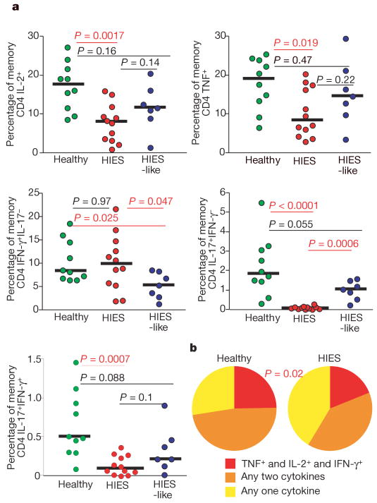
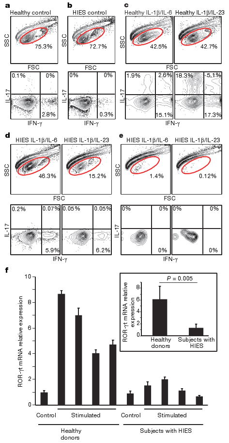
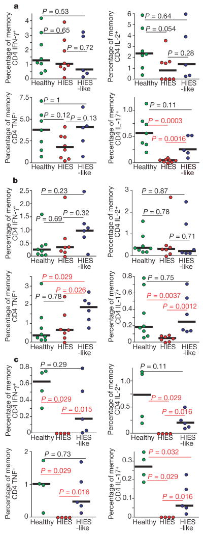
The autosomal dominant hyper-IgE syndrome (HIES, 'Job's syndrome') is characterized by recurrent and often severe pulmonary infections, pneumatoceles, eczema, staphylococcal abscesses, mucocutaneous candidiasis, and abnormalities of bone and connective tissue. Mutations presumed to underlie HIES have recently been identified in stat3, the gene encoding STAT3 (signal transducer and activator of transcription 3) (refs 3, 4). Although impaired production of interferon-gamma and tumour-necrosis factor by T cells, diminished memory T-cell populations, decreased delayed-type-hypersensitivity responses and decreased in vitro lymphoproliferation in response to specific antigens have variably been described, specific immunological abnormalities that can explain the unique susceptibility to particular infections seen in HIES have not yet been defined. Here we show that interleukin (IL)-17 production by T cells is absent in HIES individuals. We observed that ex vivo T cells from subjects with HIES failed to produce IL-17, but not IL-2, tumour-necrosis factor or interferon-gamma, on mitogenic stimulation with staphylococcal enterotoxin B or on antigenic stimulation with Candida albicans or streptokinase. Purified naive T cells were unable to differentiate into IL-17-producing (T(H)17) T helper cells in vitro and had lower expression of retinoid-related orphan receptor (ROR)-gammat, which is consistent with a crucial role for STAT3 signalling in the generation of T(H)17 cells. T(H)17 cells have emerged as an important subset of helper T cells that are believed to be critical in the clearance of fungal and extracellular bacterial infections. Thus, our data suggest that the inability to produce T(H)17 cells is a mechanism underlying the susceptibility to the recurrent infections commonly seen in HIES.


Figures

Figure 1
Figure 1. Lack of IL-17 production in SEB-stimulated PBMCs from HIES patients
a, PBMCs from cohorts of HIES patients with stat3 mutations (red circles), HIES-like patients (blue circles) and healthy individuals (green circles) were stimulated overnight with SEB in the presence of brefeldin A and stained as described in Methods. The frequency of memory CD4 T cells (gated as described in Methods) that produced IFN-γ, IL-2, TNF or IL-17 was then assessed. b, Memory CD4 T cells that produced IFN-γ were analysed for their ability to co-produce IL-2 and TNF by using SPICE (Simplified Presentation of Incredibly Complex Evaluations) as described in Methods. Statistical significance was determined with the Mann–Whitney test. Significant P values are shown in red, and median values with horizontal bars.
Figure 2
Figure 2. Failure of TH17 generation from naive cells of patients with HIES
A total of 50,000 naive T cells from healthy controls and patients with HIES were stimulated with anti-CD2/CD3/CD28 microbeads and the indicated cytokines as described in Methods. a–e, Dot plots of intracellular cytokine staining from healthy control (a) and HIES (b) cells stimulated without any cytokines, and from control subjects (c) and subjects with HIES (d, e) stimulated in the presence of IL-6 and IL-1β, or with IL-23 and IL-1β. e, Representative forward and side scatter plots of HIES cells after stimulation for 12 days show the poor recovery after stimulation with either IL-6 and IL-1β, or IL-23 and IL-1β. d, The one subject sample that had good cellular recovery. f, Naive CD4 T cells from four healthy donors and four subjects with HIES were freshly lysed or stimulated with anti-CD3 and anti-CD28 in the presence of IL-23 for 48h. ROR-γt mRNA expression was detected by quantitative RT–PCR. Values shown are relative expression levels of triplicate samples (means and s.d.). Inset: average expression levels for the four healthy donors compared with those for the four subjects with HIES. Statistical significance was determined with a two-tailed unpaired Student t-test.
Figure 3
Figure 3. Lack of IL-17 production despite antigen-specific IFN-γ, IL-2 and TNF production from subjects with HIES
PBMCs from cohorts of healthy subjects (green circles) HIES-like subjects (blue circles) and individuals with HIES (red circles) were stimulated overnight with Candida albicans (a), streptococcal kinase (b) or Staphylococcus aureus (c) in the presence of brefeldin A, then stained as described in Methods. The production of IFN-γ, IL-2, TNF or IL-17 by memory CD4 T cells (gated as described in Methods) in response to individual antigen stimulation is shown. Statistical significance was determined with the Mann–Whitney test. Significant P values are shown in red, and median values with horizontal bars.

References

    1. Buckley RH, Wray BB, Belmaker EZ. Extreme hyperimmunoglobulinemia E and undue susceptibility to infection. Pediatrics. 1972;49:59–70. - PubMed
    1. Grimbacher B, et al. Hyper-IgE syndrome with recurrent infections—an autosomal dominant multisystem disorder. N Engl J Med. 1999;340:692–702. - PubMed
    1. Minegishi Y, et al. Dominant-negative mutations in the DNA-binding domain of STAT3 cause hyper-IgE syndrome. Nature. 2007;448:1058–1062. - PubMed
    1. Holland SM, et al. STAT3 mutations in the Hyper-IgE syndrome. N Engl J Med. 2007;357:1608–1619. - PubMed
    1. Del Prete G, et al. Defective in vitro production of γ-interferon and tumor necrosis factor-α by circulating T cells from patients with the hyper-immunoglobulin E syndrome. J Clin Invest. 1989;84:1830–1835. - PMC - PubMed

Publication types

MeSH terms

LinkOut - more resources