. 2008 Aug;128(8):1950-5.
doi: 10.1038/jid.2008.52. Epub 2008 Mar 13.

Langerhans cells are not required for efficient skin graft rejection

Affiliations

Langerhans cells are not required for efficient skin graft rejection

Jagdeep S Obhrai et al. J Invest Dermatol. 2008 Aug.

Abstract

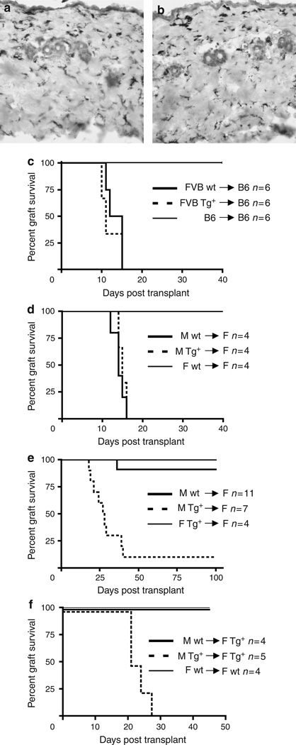
The mechanism of skin allograft rejection has been thought to require presentation of graft antigen by resident epidermal Langerhans cells (LCs). We have previously engineered mice that have a selective and constitutive absence of epidermal LCs. By using donor skin from these LC-deficient mice, we show that LCs are not required for rejection of major (FVB --> B6) or minor (H-Y, male --> female on B6 background) antigen-mismatched skin grafts. On the FVB background, where H-Y mismatched grafts are normally maintained indefinitely, grafts lacking LCs are efficiently rejected. Thus, LCs in the donor graft are required for long-term skin engraftment, which supports a regulatory role for LCs in skin graft acceptance.


Conflict of interest statement

CONFLICT OF INTEREST

The authors state no conflict of interest.

Figures

Figure 1
Figure 1. LCs are required for skin graft acceptance, but not rejection
Partial thickness grafts from FVB Tg+ (a) or control (b) mice were stained with for MHC II and counterstained with hematoxylin. Original magnification × 200. (c) Grafts harvested from wt (solid line) and Tg+ (broken line) FVB (H-2q) mice and grafted onto fully mismatched B6 (H-2b) mice. Both Tg+ and wt grafts were rejected with similar kinetics. Control syngeneic B6 grafts (thin line) were accepted indefinitely (> 100 days). (d) Skin grafts harvested from wt or Tg+ B6 males and transplanted onto B6 littermate females were rejected with similar kinetics. Control B6 female skin was accepted indefinitely. (e) Wt or Tg+ FVB male skin was placed on FVB littermate females. Tg+ grafts lacking LCs were rejected much more frequently than wt skin (P<0.001). Female Tg+ skin placed on littermate wt females was accepted indefinitely. (f) Wt or Tg+ FVB male skin was placed on Tg+ FVB littermate females. Tg+ grafts lacking LCs were rejected much more frequently than wt skin (P<0.001). Control female wt skin placed on littermate wt females was accepted indefinitely.
Figure 2
Figure 2. CD8+ cells infiltrate both wt and Tg+ FVB H-Y grafts
Graft tissue was harvested from FVB females 40 days after grafting with male wt (left panels) or Tg+ (right panels) skin. Sequential transverse sections were stained with H&E (hematoxylin and eosin) (top panels), anti-CD4 (middle panels), or anti-CD8 (bottom panels). Grafts from three mice in each group were examined and representative sections are shown. Original magnification × 100.
Figure 3
Figure 3. No differences in T-cell phenotype in Wt and Tg+ FVB H-Y graft recipients
Cells were harvested from skin-draining lymph nodes of FVB female 14 days after grafting with male wt (circle) and Tg+ (triangle) skin. Cells were stimulated ex vivo for 3 hours with PMA, and ionomycin in the presence of IL-2. Cytokine expression was measured by intracellular flow cytometry. There was no significant difference in the percentages of CD4+ (a) or CD8+ (b) T cells expressing IFN-γ, IL-2, or IL-4 in recipients grafted with Tg+ or wt skin. (c) Day 14 after transplantation of skin grafts from FVB wt or Tg+ males, the percentage of CD4+CD25+Foxp3+ cells was quantified by flow cytometry. There was no significant difference between the groups.

Comment in

References

    1. Banchereau J, Briere F, Caux C, Davoust J, Lebecque S, Liu YJ, et al. Immunobiology of dendritic cells. Annu Rev Immunol. 2000;18:767–811. - PubMed
    1. Barker CF, Billingham RE. The role of afferent lymphatics in the rejection of skin homografts. J Exp Med. 1968;128:197–221. - PMC - PubMed
    1. Benichou G, Valujskikh A, Heeger PS. Contributions of direct and indirect T cell alloreactivity during allograft rejection in mice. J Immunol. 1999;162:352–358. - PubMed
    1. Bennett CL, van Rijn E, Jung S, Inaba K, Steinman RM, Kapsenberg ML, et al. Inducible ablation of mouse Langerhans cells diminishes but fails to abrogate contact hypersensitivity. J Cell Biol. 2005;169:569–576. - PMC - PubMed
    1. Bursch LS, Wang L, Igyarto B, Kissenpfennig A, Malissen B, Kaplan DH, et al. Identification of a novel population of Langerin+ dendritic cells. J Exp Med. 2007;204:3147–3156. - PMC - PubMed

Publication types

MeSH terms

LinkOut - more resources