

Langerhans' cell histiocytosis of the spine in children with soft tissue extension and chemotherapy
- PMID: 18338168
- PMCID: PMC2903089
- DOI: 10.1007/s00264-008-0529-8
Langerhans' cell histiocytosis of the spine in children with soft tissue extension and chemotherapy
Abstract
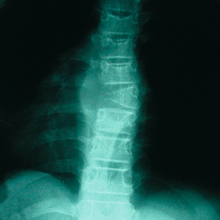
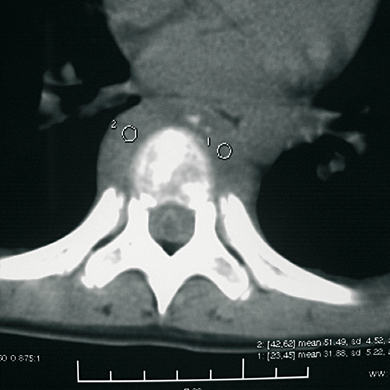
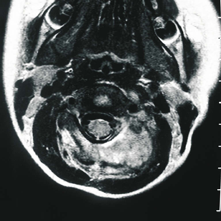
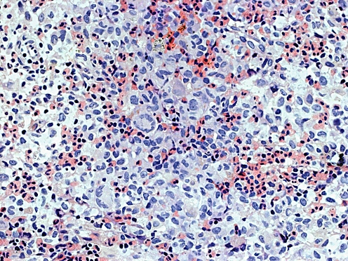
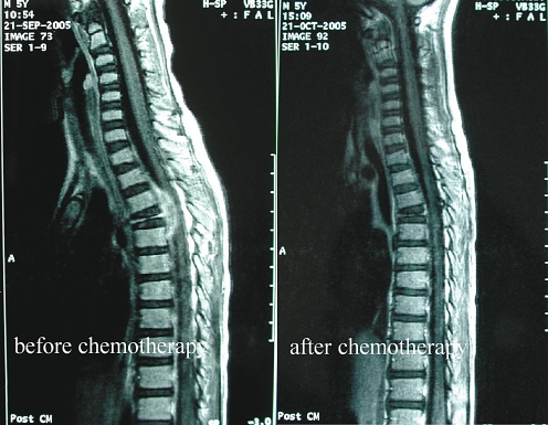
The objectives of this paper were to look into the possible incidence of obvious soft tissue extension from Langerhans' cell histiocytosis (LCH) of the spine in children and to evaluate the effects of chemotherapy for those patients. Eighteen patients with histopathological diagnosis of LCH were reviewed and nine with obvious paravertebral soft tissue extension were included in this study. Soft tissue extension was involved in the spinal canal and/or around the vertebral body in eight cases and posterior involvement was prevalent in one case. Eight patients experienced neurological symptoms. All received chemotherapy and one had surgical treatment. The mean follow-up time was 30.3 months. Soft tissue extension disappeared completely in all patients. No clinical evidence of disease was observed at the most recent follow-up. The incidence of LCH of the spine in children with obvious soft tissue extension was up to 50%. Chemotherapy is safe and effective, and surgical decompression was probably not necessary for most patients.
Objectif de l’étude : vérifier l’incidence de la pénétration tissulaire d’une histiocytose, à cellules Langerhancienne (LCH) au niveau du rachis de l’enfant et évaluer les effets de la chimiothérapie. Matériel et méthode : 18 patients avec diagnostic histologique du LCH ont été revus. 9 présentaient une invasion évidente des tissus mous et ont été inclus dans cette étude. Résultat : l’extension tissulaire a été localisée au niveau du canal médullaire ou autour du corps vertébral chez 8 patients. Cette localisation a été uniquement postérieure chez un patient. Huit présentaient des signes neurologiques et tous ont reçu de la chimiothérapie et un seul un traitement chirurgical. Le suivi moyen a été de 30,3 mois. La pénétration tissulaire a complètement disparu chez tous les patients. Il n’y a pas eu de récidive clinique de la maladie au plus récent recul. Conclusion : l’incidence dans la LCH de la pénétration tissulaire au niveau du rachis de l’enfant, est de 50%, de la chimiothérapie est un traitement sûr et efficace, il n’est pas nécessaire d’envisager une décompression chirurgicale dans la plupart des cas.
Figures
References
-
- Aster J, Kumar V. White cells, lymph nodes, spleen, and thymus. In: Cotran RS, Kumar V, Collins T, Robbins SL, editors. Robbins pathologic basis of disease. 6. Philadelphia: Saunders; 1999. pp. 644–686.
-
- Bilge T, Barut S, Yaymaci Y, et al. Solitary eosinophilic granuloma of the lumbar spine in an adult. Case report. Paraplegia. 1995;33:485–487. - PubMed
Publication types
MeSH terms
LinkOut - more resources
Full Text Sources
Medical
