Review
. 2008 Apr;7(4):327-40.
doi: 10.1016/S1474-4422(08)70060-7.

Paraneoplastic syndromes of the CNS

Affiliations
Review

Paraneoplastic syndromes of the CNS

Josep Dalmau et al. Lancet Neurol. 2008 Apr.

Abstract

Major advances in the management of paraneoplastic neurologic disorders (PND) include the detection of new antineuronal antibodies, the improved characterisation of known syndromes, the discovery of new syndromes, and the use of CT and PET to reveal the associated tumours at an early stage. In addition, the definition of useful clinical criteria has facilitated the early recognition and treatment of these disorders. In this article, we review some classic concepts about PND and recent clinical and immunological developments, focusing on paraneoplastic cerebellar degeneration, opsoclonus-myoclonus, and encephalitides affecting the limbic system.


Figures

Figure 1
Figure 1. Initial diagnostic approach to PND of the CNS
*Well characterised onconeuronal antibodies. †Lymphocytic pleocytosis, high IgG index, and oligoclonal bands with or without high protein concentration.
Figure 2
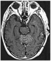
Figure 2. Early MRI findings in paraneoplastic cerebellitis
Contrast enhancement in the sulci of the cerebellar vermis in a patient with non-Hodgkin's lymphoma and subacute cerebellar degeneration in association with anti-Tr antibodies. CSF showed pleocytosis, increased protein concentration, and high titres of anti-Tr antibodies, without malignant cells. Subsequent studies showed absence of contrast enhancement and progressive atrophy (not shown).
Figure 3
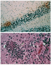
Figure 3. Inflammatory infiltrates in subacute cerebellar degeneration
CD3 T cells form clusters (A; arrows) at the level of the Purkinje cell layer. No remaining Purkinje cells are visible. Higher magnification (B) shows a neuronophagic nodule of T lymphocytes that are probably destroying a Purkinje cell. The patient had a subacute cerebellar syndrome in the context of encephalomyelitis and anti-Hu antibodies. A: ×100, immunostained for CD3 and counterstained with haematoxylin. B: ×400 haematoxylin-eosin.
Figure 4
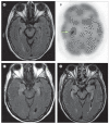
Figure 4. Neuroimaging of patients with encephalitis
A and B: patient with anti-Hu-associated sensory neuronopathy. The patient was initially treated with intravenous immunoglobulin, corticosteroids, and oral cyclophosphamide, which resulted in symptom stabilisation. MRI of the brain (A) was normal. A body fluorodeoxyglucose-PET scan that included the brain revealed an area of hypermetabolism in the right hippocampus (B). The patient never developed symptoms of limbic encephalopathy. Small-cell lung cancer was eventually identified; the patient died as a result of tumour progression. C: typical increased FLAIR signal involving the hippocampi of a patient with paraneoplastic limbic encephalitis. D: medial temporal lobe FLAIR abnormalities and atrophy in a patient with a liver transplant who developed human herpes virus 6 (HHV6) encephalitis. The clinical picture and MRI findings of HHV6 closely mimic paraneoplastic limbic encephalitis.
Figure 5
Figure 5. Antibodies associated with paraneoplastic encephalitides
A and B: antibody reactivity in a rat brain with antibodies to NR1/NR2 heteromers of the NMDA receptor (A) and anti-Hu antibodies (B). Anti-NMDA receptor antibodies produce intense immunolabelling of the neuropil of the hippocampus whereas anti-Hu antibody staining is limited to neuronal cell bodies. C: cultures of non-permeabilised live neurons immunolabelled by antibodies contained in the CSF of a patient with anti-NMDA-receptor encephalitis. D: similar cultures immunolabelled with the CSF of a patient with limbic encephalitis and cancer of the thymus. The cell-surface antigen is unknown. Treatment of the tumour and intravenous immunoglobulin resulted in complete recovery. A and B: immunoperoxidase technique ×25. C: immunofluorescence ×800. D: immunofluorescence ×400.

References

    1. Guichard MMA, Vignon G. La Polyradiculonéurite cancéreuse métastatique. Le J Médecine de Lyon. 1949:197–207. - PubMed
    1. Guichard MMA, Cabanne F, Tommasi M, Fayolle J. Polyneuropathies in cancer patients and paraneoplastic polyneuropathies. Lyon Medicale. 1956;41:309–29. - PubMed
    1. Antoine JC, Camdessanche JP. Peripheral nervous system involvement in patients with cancer. Lancet Neurol. 2007;6:75–86. - PubMed
    1. Rudnicki SA, Dalmau J. Paraneoplastic syndromes of the spinal cord, nerve, and muscle. Muscle Nerve. 2000;23:1800–18. - PubMed
    1. Rudnicki SA, Dalmau J. Paraneoplastic syndromes of the peripheral nerves. Curr Opin Neurol. 2005;18:598–603. - PubMed

Publication types

MeSH terms