Review
. 2008 Jun 1;7(6):834-48.
doi: 10.1016/j.dnarep.2008.01.017. Epub 2008 Mar 12.

Do all of the neurologic diseases in patients with DNA repair gene mutations result from the accumulation of DNA damage?

Affiliations
Review

Do all of the neurologic diseases in patients with DNA repair gene mutations result from the accumulation of DNA damage?

P J Brooks et al. DNA Repair (Amst). .

Abstract

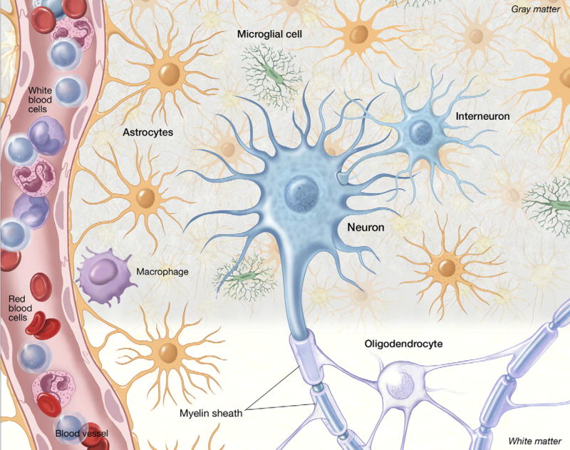
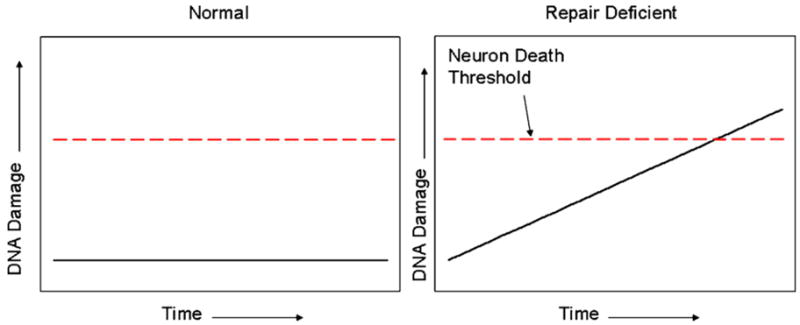
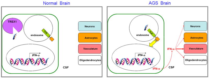
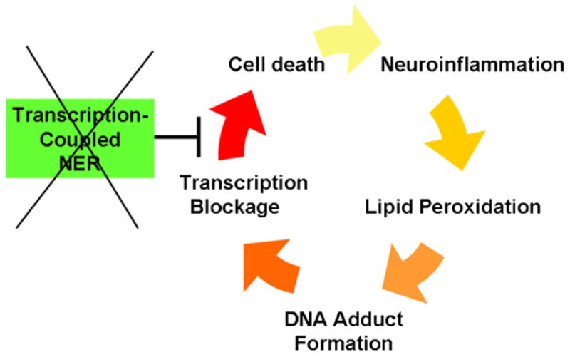
The classic model for neurodegeneration due to mutations in DNA repair genes holds that DNA damage accumulates in the absence of repair, resulting in the death of neurons. This model was originally put forth to explain the dramatic loss of neurons observed in patients with xeroderma pigmentosum neurologic disease, and is likely to be valid for other neurodegenerative diseases due to mutations in DNA repair genes. However, in trichiothiodystrophy (TTD), Aicardi-Goutières syndrome (AGS), and Cockayne syndrome (CS), abnormal myelin is the most prominent neuropathological feature. Myelin is synthesized by specific types of glial cells called oligodendrocytes. In this review, we focus on new studies that illustrate two disease mechanisms for myelin defects resulting from mutations in DNA repair genes, both of which are fundamentally different than the classic model described above. First, studies using the TTD mouse model indicate that TFIIH acts as a co-activator for thyroid hormone-dependent gene expression in the brain, and that a causative XPD mutation in TTD results in reduction of this co-activator function and a dysregulation of myelin-related gene expression. Second, in AGS, which is caused by mutations in either TREX1 or RNASEH2, recent evidence indicates that failure to degrade nucleic acids produced during S-phase triggers activation of the innate immune system, resulting in myelin defects and calcification of the brain. Strikingly, both myelin defects and brain calcification are both prominent features of CS neurologic disease. The similar neuropathology in CS and AGS seems unlikely to be due to the loss of a common DNA repair function, and based on the evidence in the literature, we propose that vascular abnormalities may be part of the mechanism that is common to both diseases. In summary, while the classic DNA damage accumulation model is applicable to the neuronal death due to defective DNA repair, the myelination defects and brain calcification seem to be better explained by quite different mechanisms. We discuss the implications of these different disease mechanisms for the rational development of treatments and therapies.


Figures

Fig. 1
Fig. 1
A drawing of the different cell types in the brain to show their anatomical relationships and relative numerical proportions. In addition to the major cell types which are discussed in the text, and marked on the figure, other notable points are the wrapping of myelin produced by oligodendrocytes around the axons of neurons, and the end-feet of the astrocytes wrapping around the blood vessel which in association with vascular endothelial cells, form the blood–brain barrier. The relative sizes of the different cell types are not drawn to scale.
Fig. 2
Fig. 2
Purkinje neurons in the mouse and human cerebellum. (Top row) (A–C) Sections through the mouse cerebellum stained with propidium iodide (PI), a nucleic acid stain, illustrating the different layers of the cerebellar cortex; ML, molecular layer; PL, Purkinje layer; GL, granule layer. (B) Same section stained with calbindin (Calb), a marker for Purkinje neurons. Panel (C) is a merged image of (A) and (B). Panel (D) is a detail from (C) to illustrate the dendritic trees of the Purkinje neurons. (Bottom row) (A) low-power image of a section through the human cerebellum (case 1465, see [35]) showing a single Purkinje neuron (arrow), adjacent to the granule layer. Note the quantitative difference between the granule neurons (in the GL, below the dotted line) relative to the single Purkinje neuron. Green staining is topoisomerase I (Top1), counterstained with PI. (B–D) Higher magnification of the Purkinje neuron shown in (A), stained with PI (B), Top1 (C) and a merged image (D). In (B), note that the nucleolus of the Purkinje neuron (arrow) is comparable in size to the entire nucleus of the granule neurons shown below. Also note the much greater intensity for Top1 staining in the nucleus of the Purkinje neuron compared to the granule neurons. For further information, see [35]. Images produced by Tracy Gilman (mouse) and Sarah Calkins (human). The conditions used for PI staining and image acquisition were substantially different between the mouse and human material, and therefore the mouse and human images are not directly comparable with each other.
Fig. 3
Fig. 3
The accumulated damage model of neurodegeneration due to defective DNA repair. For explanation see text. For the neurodegenerative diseases in Table 1, the time necessary for accumulated DNA damage to reach the neuronal death threshold is likely to be several years, if not longer. This may explain why the Atm−/−, Xpa−/− and Aptx−/− mice do not accurately reproduce the progressive neurodegeneration observed in the human patients; the 2-year lifespan of a mouse is simply not long enough to allow sufficient damage to accumulate (see also [10]).
Fig. 4
Fig. 4
A speculative working model for AGS neurologic disease, incorporating the proposals [52] and data from AGS cells [66], as well as the neuropathological evidence from AGS patients [19] and the IFN-α transgenic mice [66,68]. In one or more cell types in the normal brain, most likely microglia or macrophages, ssDNA resulting from DNA replication is produced, but is degraded by TREX1. In the AGS brain, ssDNA enters the endosome due to the absence of TREX1, where it activates a TLR. TLR activation results in signal transduction to the nucleus (yellow arrow), increasing the expression of the IFN-α gene (as well as other genes, not shown for clarity). This results in the synthesis and secretion of IFN-α into the CSF, where it can act on the other brain cell types. Effects in IFN-α in the vasculature appear to be of particular importance, and in human patients, dysmyelination is also prominent [19], indicating effects on oligodendrocytes. Activation of astrocytes and functional changes in neurons are also observed in the IFN-α overexpressing mice [66,68].
Fig. 5
Fig. 5
A speculative role for TC-NER in preventing a neuroinflammatory cycle of dysmyelination and calcification of the brain in CS. Inflammation results in lipid peroxidation and transcription-blocking DNA damage, which is repaired by TC-NER. CS gene mutations inactivate TC-NER, resulting in cell death, and in turn more inflammation from phagocytic cells. Under these conditions, the transcriptional defects in CS cells under conditions of DNA damage [78,79] may also play a role.

References

    1. Friedberg E, Walker G, Siede W, Wood R, Schultz R, Ellenberger T. DNA Repair and Mutagenesis. ASM Press; Washington, DC: 2005.
    1. Kandel E, Schwatrz J, Jessel T. Principles of Neural Science. Elsevier; New York: 1991.
    1. Ledoux SP, Shen CC, Grishko VI, Fields PA, Gard AL, Wilson GL. Glial cell-specific differences in response to alkylation damage. Glia. 1998;24:304–312. - PubMed
    1. Hollensworth SB, Shen C, Sim JE, Spitz DR, Wilson GL, LeDoux SP. Glial cell type-specific responses to menadione-induced oxidative stress. Free Radic Biol Med. 2000;28:1161–1174. - PubMed
    1. Guillemin GJ, Brew BJ. Microglia, macrophages, perivascular macrophages, and pericytes: a review of function and identification. J Leukoc Biol. 2004;75:388–397. - PubMed

Substances