. 2008 May 16;283(20):14053-62.
doi: 10.1074/jbc.M708974200. Epub 2008 Mar 13.

Structures of rhodopsin kinase in different ligand states reveal key elements involved in G protein-coupled receptor kinase activation

Affiliations

Structures of rhodopsin kinase in different ligand states reveal key elements involved in G protein-coupled receptor kinase activation

Puja Singh et al. J Biol Chem. .

Abstract

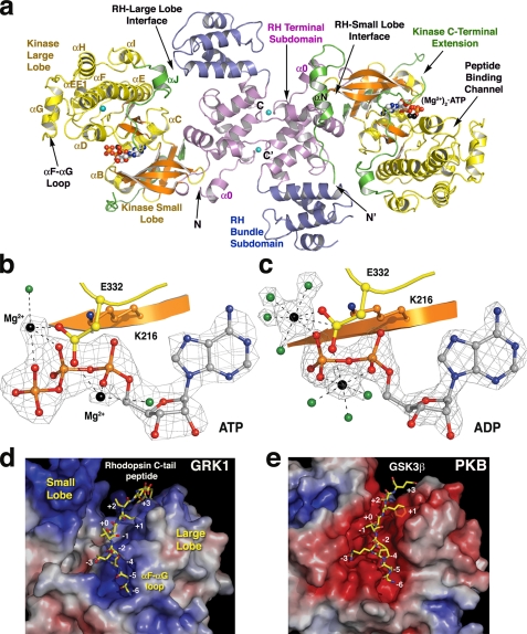
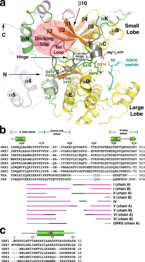
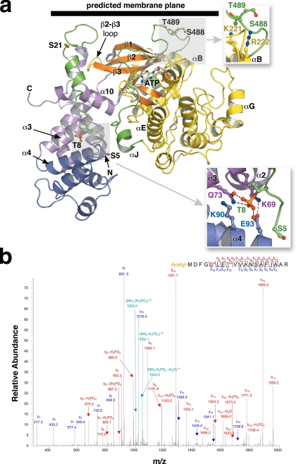
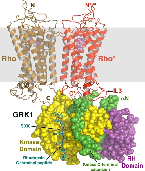
G protein-coupled receptor (GPCR) kinases (GRKs) phosphorylate activated heptahelical receptors, leading to their uncoupling from G proteins. Here we report six crystal structures of rhodopsin kinase (GRK1), revealing not only three distinct nucleotide-binding states of a GRK but also two key structural elements believed to be involved in the recognition of activated GPCRs. The first is the C-terminal extension of the kinase domain, which was observed in all nucleotide-bound GRK1 structures. The second is residues 5-30 of the N terminus, observed in one of the GRK1.(Mg2+)2.ATP structures. The N terminus was also clearly phosphorylated, leading to the identification of two novel phosphorylation sites by mass spectral analysis. Co-localization of the N terminus and the C-terminal extension near the hinge of the kinase domain suggests that activated GPCRs stimulate kinase activity by binding to this region to facilitate full closure of the kinase domain.


Figures

FIGURE 1.
FIGURE 1.
Overview of GRK1 and its active site. a, GRK1535-His6 crystallized as a homodimer using a conserved interface of the RH domain in all the crystal forms. Shown is the most complete structure, that of crystal form I. The RH terminal subdomain is colored magenta (helices α0-3 and α8-11), and the bundle subdomain (helices α4-α7) is slate blue. The small lobe of the kinase domain (yellow) is composed of six β-strands (orange) and two α-helices (αB and αC), whereas the large lobe is primarily α-helical. The ligand (Mg2+)2·ATP is drawn as spheres. Magnesium atoms are colored black, carbons are white, nitrogens are blue, oxygens are red, phosphates are orange, and chloride ions are cyan. The extreme N-terminal region and the C-terminal extension of the kinase domain are green. b, substrate complex of GRK1. Shown is a σA-weighted |Fo| - |Fc| omit map contoured at 4 σ, wherein ATP, Mg2+, and associated waters (green) were excluded from refinement (crystal form I and chain B). Lys216 (β1 sheet, orange carbons) coordinates the α- and β-phosphates. Glu332 (yellow carbons) coordinates both Mg2+ atoms. c, product complex of GRK1. Shown is a σA-weighted |Fo| - |Fc| omit map contoured at 5 σ, wherein ADP, Mg2+, and associated waters were excluded from refinement (crystal form IV). d, the peptide-binding channel of GRK1. The molecular surface of GRK1 is colored by its electrostatic potential from -7 (red, acidic) to +7 (blue, basic) kT/e-. The channel has a strikingly basic character, explaining why GRK1 prefers acidic substrates (48, 49) and how it can phosphorylate multiple closely spaced Ser and Thr residues at the C terminus of Rho*. The channel is also wider in GRK1 than in nucleotide-bound PKB (e), reflecting the more open conformation of the GRK1 kinase domain of GRK1. As a result, the phosphoacceptor oxygen of the modeled peptide is >4 Å from the γ-phosphate of ATP, which is too far for covalent chemistry to occur. A model of residues 332-345 from the C terminus of Rho*, is shown as a stick model docked to the large lobe with Ser338 in position to be phosphorylated (position “+0”). Residues in the αF-αG loop of the large lobe appear to obstruct the N-terminal end of the peptide-binding site. e, the GSK3β peptide bound to PKB. The PKB kinase domain (Protein Data Bank code 1O6L) is in its closed conformation. The channel is markedly acidic, in line with a preference for basic substrates.
FIGURE 2.
FIGURE 2.
The N terminus and kinase C-terminal extension of GRK1. a, the active site regions of GRK1 and PKB. For this comparison, the coordinates of the various GRK1 crystal forms were merged to generate a composite structure of GRK1 that spans residues 5-533 (of 558), and the large lobe was rotated to generate the expected closed, active state. The kinase C-terminal extension of PKB is gray, and the GSK3β peptide bound to PKB is cyan. The tail-loop of GRKs is four residues shorter than those of either PKB or PKA, resulting in formation of a shallow canyon with the hinge of the kinase domain at the bottom. This region forms a putative receptor-docking site (transparent ellipse). b, structural alignment of the kinase C-terminal extension of GRKs with PKA and PKB. The region immediately following the active site tether is structurally divergent among GRKs, PKA, and PKB, and thus no alignment is attempted. The numbers above the alignment refer to the sequence of bovine GRK1. The different segments of the C-terminal extension that were observed in each of our crystal forms are indicated below the alignment, with the black regions corresponding to structurally heterogeneous portions in each structure that appear influenced by crystal contacts. The accession codes for the protein sequences are: GRK1, P28327; GRK2, NP_777135.1; GRK3, P26818; GRK4, AAI17321.1; GRK5, P43249; GRK6, P43250; GRK7, NP_776757.1; PKA, 1L3R; PKB, 1O6L. c, structural alignment of GRK N-terminal regions.
FIGURE 3.
FIGURE 3.
The phosphorylation sites of GRK1. a, the RH-kinase core of GRK1. The structure corresponds to that of crystal form I (with composite C-terminal extension; see Fig. 2). The Ser5, Thr8, Ser21, Ser488, and Thr489 phosphorylation sites are drawn as stick models. The expected position of the membrane plane is indicated. Top inset, the Ser488 and Thr489 phosphorylation sites correspond to the AGC kinase turn motif. Bottom inset, interaction of Thr(P)8 with the RH domain. Gln73 and Glu93 form direct hydrogen bonds, whereas Lys69 and Lys90 complement the charge of the phosphate moiety. These crystals grew at pH 4.35, and so either Glu93 or the phosphate group could be protonated. b, tandem mass spectrometry spectra of phosphopeptides from GRK1535-His6 (Pool A, pretreated with 4 mm ATP and 2 mm MgCl2). Both Ser5 and Thr8 sites were identified in a single peptide. The Ser5 site was also readily observed in endogenous GRK1, as were the previously observed phosphorylation sites at Ser21, Ser488, and Thr489 (supplemental Fig. S7).
FIGURE 4.
FIGURE 4.
Conceptual model of GRK1 docked to Rho*. The closed composite model of GRK1 (Fig. 2a) was docked with a model of an array of Rho molecules (Protein Data Bank code 1N3M) (50), of which two molecules are shown here for clarity. GRK1 is rendered as spheres, and the expected lipid bilayer plane is shown as a transparent gray box. A monomer of Rho* (red) was modeled such that its third cytoplasmic loop (IL3) lies close to the proposed receptor-docking site. Using the PKB-GSK3β structure (1O6L) as a guide, the C-terminal peptide of Rho* (carbons are colored cyan, oxygens are red, and nitrogens are blue) was modeled docked to the large lobe, as in Fig. 1d. The GRK1 active site would have easy access to the C-tail of Rho* or of a neighboring unactivated Rho (brown) in the same membrane plane, allowing high gain phosphorylation of ROS.

References

    1. Palczewski, K. (2006) Annu. Rev. Biochem. 75 743-767 - PMC - PubMed
    1. Maeda, T., Imanishi, Y., and Palczewski, K. (2003) Prog. Retin Eye Res. 22 417-434 - PubMed
    1. Arshavsky, V. Y., Lamb, T. D., and Pugh, E. N., Jr. (2002) Annu. Rev. Physiol. 64 153-187 - PubMed
    1. Doan, T., Mendez, A., Detwiler, P. B., Chen, J., and Rieke, F. (2006) Science 313 530-533 - PubMed
    1. Yamamoto, S., Sippel, K. C., Berson, E. L., and Dryja, T. P. (1997) Nat. Genet. 15 175-178 - PubMed

Publication types

Substances