Case Reports
. 2008 Jun;131(Pt 6):1630-45.
doi: 10.1093/brain/awn016. Epub 2008 Mar 12.

Post-mortem correlates of in vivo PiB-PET amyloid imaging in a typical case of Alzheimer's disease

Affiliations
Case Reports

Post-mortem correlates of in vivo PiB-PET amyloid imaging in a typical case of Alzheimer's disease

Milos D Ikonomovic et al. Brain. 2008 Jun.

Abstract

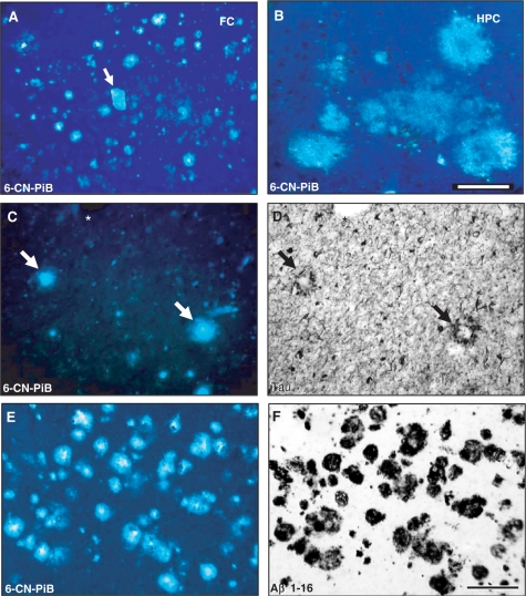
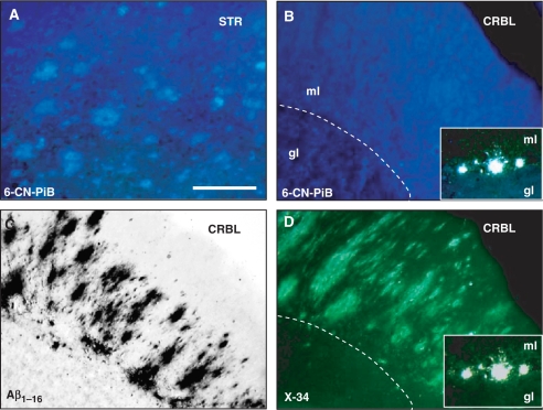
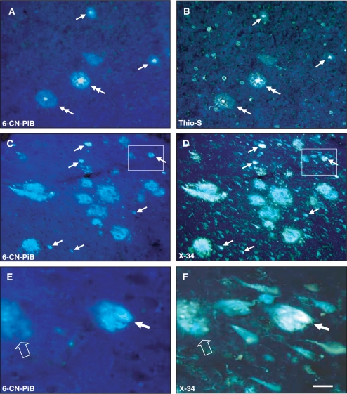
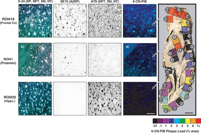
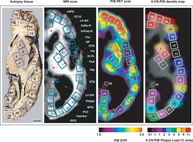
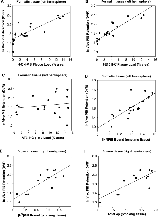
The positron emission tomography (PET) radiotracer Pittsburgh Compound-B (PiB) binds with high affinity to beta-pleated sheet aggregates of the amyloid-beta (Abeta) peptide in vitro. The in vivo retention of PiB in brains of people with Alzheimer's disease shows a regional distribution that is very similar to distribution of Abeta deposits observed post-mortem. However, the basis for regional variations in PiB binding in vivo, and the extent to which it binds to different types of Abeta-containing plaques and tau-containing neurofibrillary tangles (NFT), has not been thoroughly investigated. The present study examined 28 clinically diagnosed and autopsy-confirmed Alzheimer's disease subjects, including one Alzheimer's disease subject who had undergone PiB-PET imaging 10 months prior to death, to evaluate region- and substrate-specific binding of the highly fluorescent PiB derivative 6-CN-PiB. These data were then correlated with region-matched Abeta plaque load and peptide levels, [(3)H]PiB binding in vitro, and in vivo PET retention levels. We found that in Alzheimer's disease brain tissue sections, the preponderance of 6-CN-PiB binding is in plaques immunoreactive to either Abeta42 or Abeta40, and to vascular Abeta deposits. 6-CN-PiB labelling was most robust in compact/cored plaques in the prefrontal and temporal cortices. While diffuse plaques, including those in caudate nucleus and presubiculum, were less prominently labelled, amorphous Abeta plaques in the cerebellum were not detectable with 6-CN-PiB. Only a small subset of NFT were 6-CN-PiB positive; these resembled extracellular 'ghost' NFT. In Alzheimer's disease brain tissue homogenates, there was a direct correlation between [(3)H]PiB binding and insoluble Abeta peptide levels. In the Alzheimer's disease subject who underwent PiB-PET prior to death, in vivo PiB retention levels correlated directly with region-matched post-mortem measures of [(3)H]PiB binding, insoluble Abeta peptide levels, 6-CN-PiB- and Abeta plaque load, but not with measures of NFT. These results demonstrate, in a typical Alzheimer's disease brain, that PiB binding is highly selective for insoluble (fibrillar) Abeta deposits, and not for neurofibrillary pathology. The strong direct correlation of in vivo PiB retention with region-matched quantitative analyses of Abeta plaques in the same subject supports the validity of PiB-PET imaging as a method for in vivo evaluation of Abeta plaque burden.


Figures

Fig. 1
Fig. 1
Regional variations in 6-CN-PiB fluorescence signal in Alzheimer's disease brain tissue. In the frontal cortex (FC), 6-CN-PiB histofluorescence is seen in numerous compact/cored and diffuse plaques, and in an isolated large blood vessel with amyloid angiopathy (A, arrow). Amorphous diffuse plaques in the hippocampus (HPC; B) are 6-CN-PiB positive, but have lower fluorescence intensity compared to cored/compact 6-CN-PiB labelled plaques. 6-CN-PiB histofluorescence is prominent in cores of neuritic plaques identified by the presence of tau-immunoreactive dystrophic neurites (C and D; double labelling of the same Alzheimer's disease frontal cortex tissue section with tau IHC; asterisk denotes a tissue landmark, matched plaques are marked by arrows). 6-CN-PiB and Aβ immunoreactivity co-localize in plaques in an Alzheimer's disease frontal cortex (E, F; tissue section double labelled for 6-CN-PiB histofluorescence and Aβ immuhistochemistry with 10D5 antibody). Both markers are seen exclusively in amyloid plaques. Scale bar = 175 µm (A), 100 µm (B–F).
Fig. 2
Fig. 2
6-CN-PiB histofluorescence is detectable in cloud-like diffuse plaques in the caudate nucleus of the striatum (STR; A). In the cerebellum (CRBL), 6-CN-PiB labelling is not appreciably above background levels (B), although the adjacent tissue sections processed for Aβ IHC (C, 10D5 antibody) and X-34 (D) show fleecy diffuse plaques in the molecular layer (ml) of cerebellar cortex (dotted line in B and D approximates the Purkinje cell layer). In the Purkinje cell layer, some isolated small compact plaques are detectable with both 6-CN-PiB and X-34 (inserts in B and D; gl, granular layer). Scale bar = 150 μm (A–D), 75 µm (Inset).
Fig. 3
Fig. 3
Co-localization of 6-CN-PiB (A, C, E) with Thio-S (B) and X-34 (D, F) in plaques, but not in NFT in Alzheimer's disease temporal cortex (A, B), and hippocampus (C–F). Large cored plaques (double arrows) and small burnt out plaques (arrows) are labelled with both 6-CN-PiB and Thio-S in the temporal cortex, but Thio-S also labels numerous NFT that are not detectable with 6-CN-PiB (A, B). There is a good co-localization of 6-CN-PiB and X-34 in plaques in the hippocampus (C, D). X-34 also labels an array of neurofibrillary pathology in the hippocampus (D); only small numbers of these structures are detectable with 6-CN-PiB (C, arrows). E and F represent boxed areas in C and D, respectively, and illustrate a structure labelled with both 6-CN-PiB and X-34 (small arrow) that resembles an extracellular NFT with distinct bundles of longitudinally oriented fibrils that are loosely dispersed in the neuropil. A small diffuse plaque is also double labelled (empty arrow), while the rest of the X-34 positive NFT in the field are not detectable with 6-CN-PiB. Scale bar = 75 µm (A–D), 25 µm (E, F).
Fig. 4
Fig. 4
6-CN-PiB plaque load (% area) values, quantified in each of the 25 tissue cubes dissected from an axial post-mortem tissue block, are pseudo-colour-coded on a rainbow scale from black/purple (low% area) to red (high% area). Images of 6-CN-PiB and X-34 histofluorescence, and 6E10 and AT8 IHC, were obtained in adjacent tissue sections from tissue cube #16 (frontal cortex), tissue cube #1 (striatum) and tissue cube #25 (hippocampus), and are each representative of an area with highest concentrations of labelled elements. In all tissue cubes, 6-CN-PiB exclusively labels plaques, matching the distribution of 6E10 immunoreactive and X-34 labelled plaques, but not AT8 immunoreactive or X-34 labelled NFT, DN and NT. Abbreviations: IC = internal capsule (low background fluorescence in the white matter tracts); NFT = neurofibrillary tangles; DN = dystrophic neurites; NT = neuropil threads; Hippc = hippocampus; Cx = cortex. Scale bar = 150 µm for tissue cube #16 and #1, 75 µm for tissue cube #25 and 1 cm for tissue slab.
Fig. 5
Fig. 5
Correlation between ante-mortem PiB-PET retention levels and a post-mortem VOI-matched 6-CN-PiB plaque density map as a ‘virtual PiB scan’ of the same subject. The face of the autopsy tissue block is at the level of the anterior and posterior commisures. On the autopsy tissue (left) each of the 25 blue squares represents the face of a tissue cube that was dissected from this 1-cm formalin-fixed slab. ROIs corresponding to these 25 tissue cubes were drawn on the corresponding four 0.24-cm slices of this same patient's MRI scan (second from the left; taken at the time of the PiB scan 10 months before death). These same ROIs were then transferred to the co-registered PiB-PET scan (third from left) for quantification of the in vivo PiB retention. Some tissue distortion post-mortem is evident, due to the collapse of the ventricles. On the 6-CN-PiB density map (right), the data from the post-mortem regional 6-CN-PiB plaque load analysis (see Fig. 4) were digitally blurred to create the ‘virtual PiB scan’, shown overlying the contour of the MRI scan for ROI comparison purposes. This post-mortem 6-CN-PiB plaque density map closely reflects the distribution of PiB retention levels in the PiB-PET scan recorded ante-mortem in the same person. Abbreviations: InfFG = inferior frontal gyrus; CC:G = corpus callosum: genu; LV:AH = lateral ventricle: anterior horn; CaNu:H = caudate nucleus: head; IntCap:G = internal capsule: genu; Put = putamen; GP = globus pallidus; STG = superior temporal gyrus; Ins = insular cortex; IC:PL = internal capsule: posterior limb; Thal = thalamus; Hip = hippocampus; STG = superior temporal gyrus; LV:OH = lateral ventricle: occipital horn; PHipG = parahippocampal gyrus; MTG = medial temporal gyrus; PVC = primary visual cortex; CalcSul = calcarine sulcus; OCG = occipital gyrus. Scale bar = 1 cm.
Fig. 6
Fig. 6
Correlations of the in vivo PiB DVR values from the 19 VOIs (six of the 25 VOIs had <70% tissue and were not included in the analysis, see Methods section) on the PET scan with post-mortem quantifications of 6-CN-PiB (A) or Aβ (B) plaque load, neurofibrillary load (C) and [3H]PiB binding (D) in the corresponding 19 tissue cubes from the formalin fixed left hemisphere. In vivo PiB-PET retention (DVR) levels correlate directly with regional post-mortem quantitations of 6-CN-PiB (A) and 6E10 (B) plaque loads as well as in vitro PiB binding (D), but not with AT8 neurofibrillary load (C) in formalin-fixed tissue from the left hemisphere. In fresh-frozen tissue from the right hemisphere of the same Alzheimer's disease subject, there is a significant correlation of both in vitro PiB binding (E) and total insoluble Aβ (Aβ1–40 + Aβ1–42) peptide levels (F) with in vivo PiB-PET retention (DVR) levels.

References

    1. Consensus report of the Working Group on “Molecular and Biochemical Markers of Alzheimer's Disease”. The Ronald and Nancy Reagan Research Institute of the Alzheimer's Association and the National Institute on Aging Working Group. Neurobiol Aging. 1998;19:109–16. - PubMed
    1. Bacskai BJ, Frosch MP, Freeman SH, Raymond SB, Augustinack JC, Johnson KA, et al. Molecular imaging with PiB confirmed at autopsy: a case report. Arch Neurol. 2007;64:431–4. - PubMed
    1. Braak H, Braak E. Neuropathological stageing of Alzheimer-related changes. Acta Neuropath. 1991a;82:239–59. - PubMed
    1. Braak H, Braak E. Neuropathological staging of Alzheimer's disease. Acta Neuropath. 1991b;82:239–59. - PubMed
    1. DeKosky ST, Abrahamson EE, Ciallella JR, Paljug WR, Wisniewski SR, Clark RSB, et al. Association of increased cortical soluble abeta42 levels with diffuse plaques after severe brain injury in humans. Arch Neurol. 2007;64:541–44. - PubMed

Publication types

MeSH terms