. 2008 Mar 25;105(12):4808-13.
doi: 10.1073/pnas.0710907105. Epub 2008 Mar 13.

Role of the Akt pathway in mRNA translation of interferon-stimulated genes

Affiliations

Role of the Akt pathway in mRNA translation of interferon-stimulated genes

Surinder Kaur et al. Proc Natl Acad Sci U S A. .

Abstract

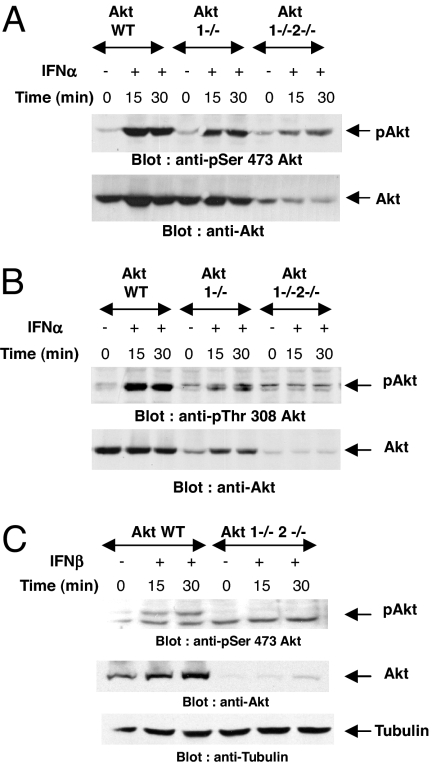
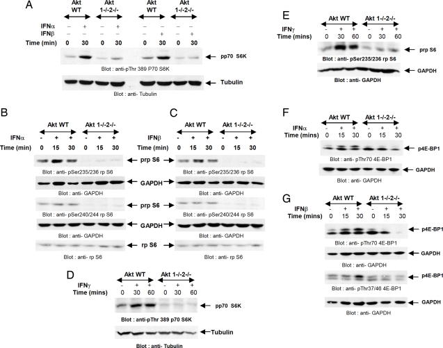
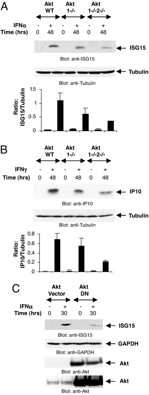
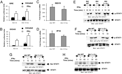
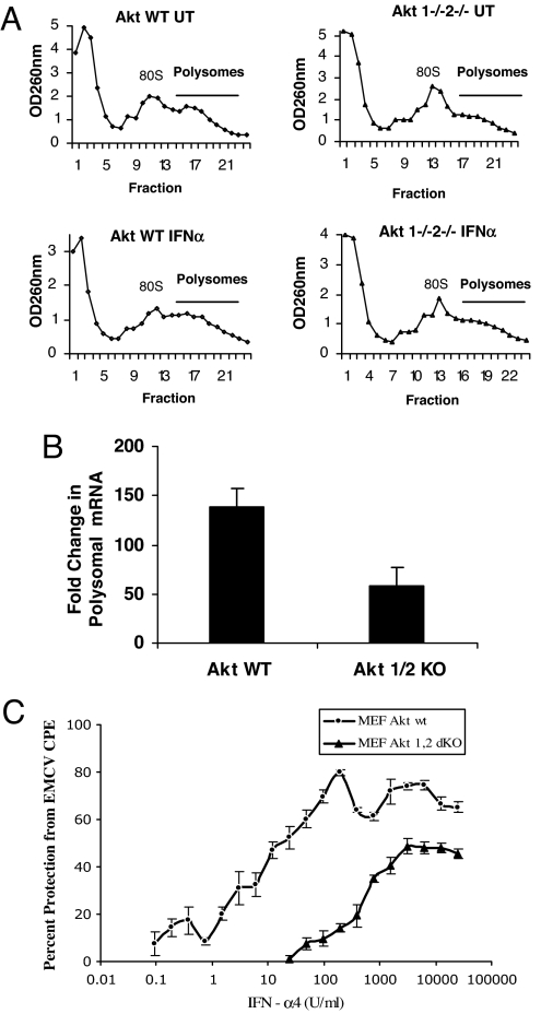
Multiple signaling pathways are engaged by the type I and II IFN receptors, but their specific roles and possible coordination in the generation of IFN-mediated biological responses remain unknown. We provide evidence that activation of Akt kinases is required for IFN-inducible engagement of the mTOR/p70 S6 kinase pathway. Our data establish that Akt activity is essential for up-regulation of key IFN-alpha- and IFN-gamma-inducible proteins, which have important functional consequences in the induction of IFN responses. Such effects of the Akt pathway are unrelated to regulatory activities on IFN-dependent STAT phosphorylation/activation or transcriptional regulation. By contrast, they reflect regulatory activities on mRNA translation via direct control of the mTOR pathway. In studies using Akt1 and Akt2 double knockout cells, we found that the absence of Akt kinases results in dramatic reduction in IFN-induced antiviral responses, establishing a critical role of the Akt pathway in IFN signaling. Thus, activation of the Akt pathway by the IFN receptors complements the function of IFN-activated JAK-STAT pathways, by allowing mRNA translation of IFN-stimulated genes and, ultimately, the induction of the biological effects of IFNs.


Conflict of interest statement

The authors declare no conflict of interest.

Figures

Fig. 1.
Fig. 1.
Type I IFN-dependent phosphorylation/activation of Akt. (A and B) Akt1+/+2+/+, Akt1−/−2+/+, and Akt1−/−2−/− MEFs were treated with mouse IFN-α for the indicated times. Equal protein aliquots were processed for immunoblotting with antibodies against phosphorylated forms of Akt on Ser-473 (A) or Thr-308 (B). Respective blots were stripped and reprobed with a pan anti-Akt antibody as indicated. (C) Akt1+/+2+/+ and Akt1−/−2−/− MEFs were treated with mouse IFN-β for the indicated times. (Top) Equal protein aliquots were immunoblotted with an antibody against the phosphorylated form of Akt on Ser-473. (Middle) Equal protein aliquots from the same experiment were resolved on separate gels and probed with an anti-Akt antibody. (Bottom) The blot shown in Top was reprobed with an anti-tubulin antibody to control for protein loading.
Fig. 2.
Fig. 2.
Requirement of Akt1 and Akt2 for type I and II IFN-dependent engagement of mTOR-regulated signaling cascades. (A) Akt1+/+2+/+ and Akt1−/−2−/− MEFs were treated with mouse IFN-α or mouse IFN-β for the indicated times. Equal protein aliquots were processed for immunoblotting with an antibody against the phosphorylated form of p70 S6 kinase on Thr-389. The same blot was reprobed with an anti-tubulin antibody. (B and C) Akt1+/+2+/+ and Akt1−/−2−/− MEFs were treated with mouse IFN-α (B) or mouse IFN-β (C) for the indicated times. Equal protein aliquots were processed for immunoblotting with antibodies against the phosphorylated forms of the S6 ribosomal protein on Ser-235/236 or Ser-240/244. The respective blots were reprobed with anti-GAPDH antibody as indicated. Equal protein aliquots from the same experiments were resolved on separate gels and immunoblotted with an antibody against rpS6. (D and E) Akt1+/+2+/+ and Akt1−/−2−/− MEFs were treated with mouse IFN-γ, and equal protein aliquots were processed for immunoblotting with an antibody against the phosphorylated form of p70 S6 kinase on Thr-389 (D Upper) or with an antibody against the phosphorylated form of rp S6 on Ser-235/236 (E Upper). Respective blots were reprobed with an anti-tubulin antibody (D Lower) or an anti-GAPDH antibody (E Lower). (F and G) Akt1+/+2+/+ and Akt1−/−2−/− MEFs were treated with mouse IFN-α (F) or mouse IFN-β (G) for the indicated times. Equal protein aliquots were processed for immunoblotting with antibodies against the phosphorylated forms of 4EBP1 on Thr-70 or Thr-37/46, as indicated. The same blots were reprobed with GAPDH to control for protein loading.
Fig. 3.
Fig. 3.
Akt is required for IFN-dependent ISG15 and CXCL10 protein expression. (A) Akt1+/+2+/+, Akt1−/−2+/+, and Akt1−/−2−/− MEFs were treated with mouse IFN-α as indicated. Equal protein aliquots were processed for immunoblotting with an anti-mouse ISG15 antibody. The same blot was reprobed with an anti-tubulin antibody as indicated. The signals for ISG15 and tubulin from three independent experiments (including the one shown) were quantitated by densitometry, and the intensity of ISG15 relative to tubulin was calculated. Data are expressed as means of ratios of ISG15/tubulin ± SE for each experimental condition. (B) Akt MEFs were treated with mouse IFN-γ as indicated. Equal protein aliquots were processed for immunoblotting with an anti-mouse IP10 antibody. Same blot was reprobed with anti-tubulin antibody as indicated. The signals for IP10 and tubulin from two independent experiments (including the one shown) were quantitated by densitometry, and the intensity of IP10 relative to tubulin was calculated. Data are expressed as means of ratios of IP10/tubulin ± SE for each experimental condition. (C) U20S cells were transfected with either the control empty vector or a dominant-negative Akt mutant and treated with human IFN-α as indicated. Equal protein aliquots were processed for immunoblotting with anti-ISG15 antibody. The blot was stripped and probed with anti-GAPDH antibody. Lysates from the same experiment were resolved separately and immunoblotted with an anti-Akt antibody. Also shown is longer exposure of the same blot to demonstrate the presence of endogenous Akt protein.
Fig. 4.
Fig. 4.
IFN-dependent gene transcription and STAT activation is intact in the absence of Akt. (A and B) Akt1+/+2+/+ or Akt1−/−2−/− MEFs were transfected with a β-galactosidase expression vector and either ISRE (A) or GAS (B) luciferase plasmids. Triplicate cultures were either left untreated or treated with IFN-α (A) or IFN-γ (B) for 6 h, and luciferase reporter assays were performed. The data are expressed as relative luciferase units for each condition, normalized for β-galactosidase activities, and represent means ± SE. Values of five independent experiments are shown for A, and values of four independent experiments are shown for B. (C and D) Akt1+/+2+/+ or Akt1−/−2−/− MEFs were incubated in presence or absence of IFN-α (C) or IFN-γ (D). Expression of mRNA for Isg15 or Cxcl10 genes was evaluated by quantitative RT-PCR (Taqman). GAPDH was used for normalization. Data are expressed as fold increase over untreated sample and represent means ± SE of five experiments. (E and F) Akt MEFs were treated with mouse IFN-α (E) or IFN-γ (F) as indicated. Equal protein aliquots were processed for immunoblotting with an antibody against the phosphorylated form of Stat1 on Tyr-701 (Upper), and blots were stripped and reprobed with an anti-Stat1 antibody (Lower). (G and H) Akt MEFs were treated with mouse IFN-α (G) or IFN-γ (H) for indicated times. Equal protein aliquots were processed for immunoblotting with an antibody against the phosphorylated form of Stat1 on Ser-727 (Upper). The same blots were stripped and reprobed with an anti-Stat1 antibody (Lower).
Fig. 5.
Fig. 5.
Engagement of Akt is essential for IFN-dependent mRNA translation and generation of antiviral responses. (A) Akt1+/+2+/+ and Akt1−/−2−/− MEFs were either left untreated or treated with mIFN-α. Cell lysates were separated on 10–50% sucrose gradient, and OD at 260 nm was recorded. The OD260 is shown as a function of gradient depth for each treatment. (B) Polysomal fractions were collected as indicated in A, and RNA was isolated. Subsequently, quantitative real-time RT-PCR assays to determine Isg15 mRNA expression in the polysomal fractions was carried out, using GAPDH for normalization. Data are expressed as fold increase over IFN-α-untreated samples and represent means ± SE of three independent experiments. Paired t test analysis comparing induction of Isg15 mRNA in polysomal fractions of Akt1−/−2−/− MEFs versus Akt1+/+2+/+ MEFs showed a paired P value = 0.0087. (C) Akt1+/+2+/+ and Akt1−/−2−/− MEFs were incubated in triplicate with the indicated doses of mouse IFN-α. The cells were subsequently challenged with EMCV, and the cytopathic effects (CPE) were quantified 4 days later. Data are expressed as percent protection from CPE of EMCV.

References

    1. Pestka S, Langer JA, Zoon KC, Samuel CE. Interferons and their actions. Annu Rev Biochem. 1987;56:727–777. - PubMed
    1. Darnell JE, Jr, Kerr IM, Stark GR. Jak-STAT pathways and transcriptional activation in response to IFNs and other extracellular signaling proteins. Science. 1994;264:1415–1420. - PubMed
    1. Stark GR, Kerr IM, Williams BR, Silverman RH, Schreiber RD. How cells respond to interferons. Annu Rev Biochem. 1998;67:227–264. - PubMed
    1. Platanias LC, Fish EN. Signaling pathways activated by interferons. Exp Hematol. 1999;27:1583–1592. - PubMed
    1. Platanias LC. Mechanisms of type I and type II interferon-mediated signaling. Nat Rev Immunol. 2005;5:375–386. - PubMed

Publication types

MeSH terms

LinkOut - more resources