. 2008 Jun;325(3):801-8.
doi: 10.1124/jpet.108.136721. Epub 2008 Mar 13.

New role of resistin in lipopolysaccharide-induced liver damage in mice

Affiliations

New role of resistin in lipopolysaccharide-induced liver damage in mice

Juliane I Beier et al. J Pharmacol Exp Ther. 2008 Jun.

Abstract

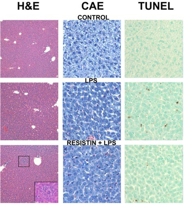
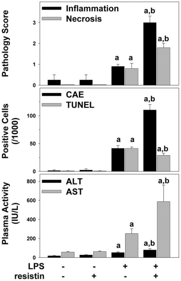
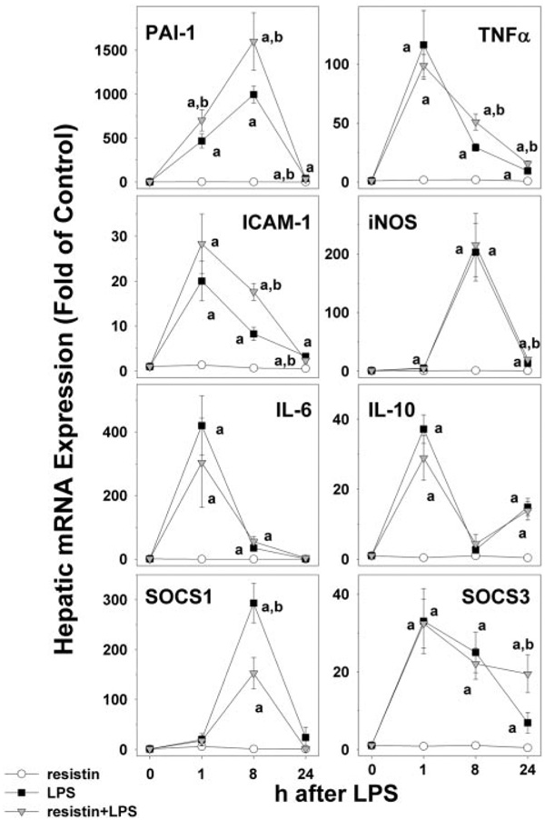
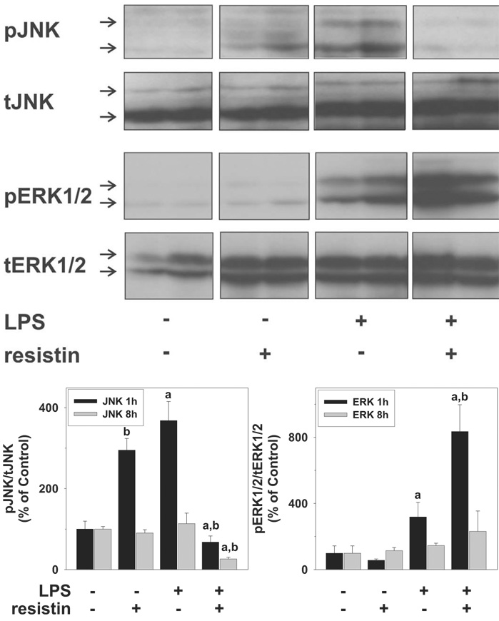
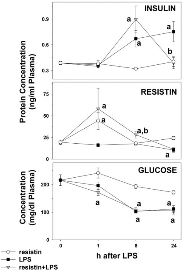
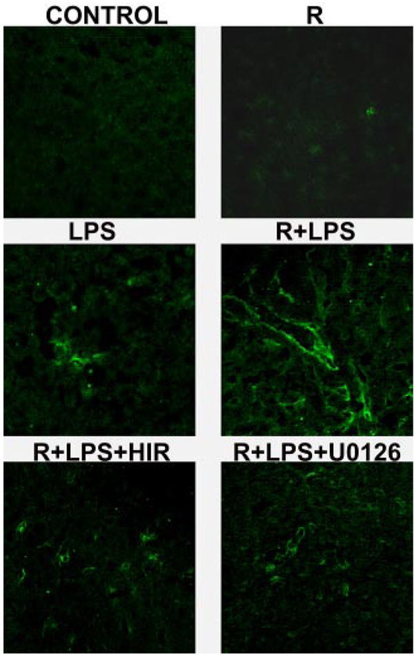
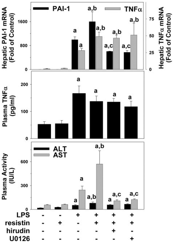
Studies in rodents suggest that the adipocytokine resistin causes insulin resistance via impairing normal insulin signaling. However, in humans, resistin may play a more important role in inflammation than in insulin resistance. Whether resistin contributes to inflammation in rodents is unclear. Therefore, the purpose of the present study was to determine the effect of resistin exposure on the basal and stimulated [lipopolysaccharide (LPS)] inflammatory response in mouse liver in vivo. Resistin alone had no major effects on hepatic expression of insulin-responsive genes, either in the presence or absence of LPS. Although it had no effect alone, resistin significantly enhanced hepatic inflammation and necrosis caused by LPS. Resistin increased expression of proinflammatory genes, e.g., plasminogen activator inhibitor (PAI)-1, and activity of mitogen-activated protein (MAP) kinase, extracellular signal-regulated kinase 1/2, caused by LPS, but had little effect on anti-inflammatory gene expression. Resistin also enhanced fibrin deposition (an index of hemostasis) caused by LPS. The increase in PAI-1 expression, fibrin deposition, and liver damage caused by LPS + resistin was almost completely prevented either by inhibiting the coagulation cascade, hirudin, or by blocking MAP kinase signaling, U0126 [1,4-diamino-2,3-dicyano-1,4-bis(2-aminophenylthio) butadiene], indicating that these pathways play a causal role in observed enhanced liver damage caused by resistin. Taken together, the augmentation of LPS-induced liver damage caused by resistin seems to involve, at least in part, up-regulation of hepatic inflammation via mechanisms most likely involving the coagulation cascade and fibrin accumulation. These data also suggest that resistin may have proinflammatory roles in mouse liver independent of its effects on insulin signaling, analogous to previous work in humans.


Figures

Fig. 1
Fig. 1
Photomicrographs of livers 24 h after LPS and/or resistin injection. Representative photomicrographs of hematoxylin and eosin (H&E), 200× (left); CAE, 400× (center); and TUNEL, 400× (right) stains are shown. Livers from mice that received saline injections are shown to represent both saline- and resistin-treated animals. Inset, bottom left, necroinflammatory area in the resistin/LPS group.
Fig. 2
Fig. 2
Plasma transaminase levels and quantitation of histological changes 24 h after LPS and/or resistin injection. Mice were treated as described under Materials and Methods. Pathology was scored (top panel) as described under Materials and Methods. CAE-positive cells (middle, black bars) and TUNEL-positive cells (middle, gray bars) were quantitated as described under Materials and Methods. ALT (bottom, black bars) and AST (bottom, gray bars) were determined in plasma samples for the 24-h time point. Data are means ± S.E.M. (n = 4–6) and are reported as -fold of control values. a, p < 0.05 compared with the absence of LPS; b, p < 0.05 compared with the absence of resistin.
Fig. 3
Fig. 3
Effect of resistin and LPS on the expression of proinflammatory and antiinflammatory genes in mouse liver. Real-Time RT-PCR results for the 0-, 1-, 8-, and 24-h time points were normalized to β-actin. Data represent means ± S.E.M. (n = 4–6). a, p < 0.05 compared with the absence of LPS; b, p < 0.05 compared with the absence of resistin.
Fig. 4
Fig. 4
Effect of resistin and LPS on the activation of mitogen-activated protein kinases ERK1/2 and JNK in mouse liver. The top panels depict representative bands from the same blot of ERK1/2 (p42/p44) and JNK (p46/p54) of the 1-h time point, and the bottom panel summarizes densitometric analysis of the 1- and 8-h time points. Data are means ± S.E.M. (n = 4–6) and are reported as -fold of control values. a, p < 0.05 compared with the absence of LPS;b, p < 0.05 compared with the absence of resistin.
Fig. 5
Fig. 5
Effect of LPS and resistin on plasma levels of insulin and resistin. Mice received injections with resistin, resistin + LPS, or saline as described under Materials and Methods. Protein concentrations of insulin (top panel) and resistin (middle) as well as plasma glucose (bottom) were determined. Assays were performed as described under Materials and Methods. Data are means ± S.E.M. (n = 4–6). a, p < 0.05 compared with the absence of LPS; b, p < 0.05 compared with the absence of resistin.
Fig. 6
Fig. 6
Effect of resistin and LPS on fibrin deposition 24 h after injection. Representative confocal photomicrographs (400×) depicting immunofluorescent detection of hepatic fibrin are shown.
Fig. 7
Fig. 7
Effect of Hirudin and U0126 on resistin and LPS-induced PAI-1 expression and liver damage. Mice received injections with resistin, resistin + LPS, or saline as described under Materials and Methods. Realtime RT-PCR (top panel), ELISA for TNFα protein (middle), and spectrophotometric determination of ALT/AST were performed as described under Materials and Methods. Data are means ± S.E.M. (n = 4–6). PCR data are reported as -fold of control values. a, p < 0.05 compared with the absence of LPS; b, p < 0.05 compared with the absence of resistin;c, p < 0.05 compared with the absence of hirudin or U0126. PAI-1 data for control, LPS, and resistin + LPS are derived from Fig. 3 and shown here for comparison. Transaminase data for control, LPS, and resistin + LPS are derived from Fig. 2 and shown here for comparison.

Similar articles

Cited by

References

    1. Banerjee RR, Rangwala SM, Shapiro JS, Rich AS, Rhoades B, Qi Y, Wang J, Rajala MW, Pocai A, Scherer PE, et al. Regulation of fasted blood glucose by resistin. Science. 2004;303:1195–1198. - PubMed
    1. Bergheim I, Guo L, Davis MA, Lambert JC, Beier JI, Duveau I, Luyendyk JP, Roth RA, Arteel GE. Metformin prevents alcohol-induced liver injury in the mouse: critical role of plasminogen activator inhibitor-1. Gastroenterology. 2006a;130:2099–2112. - PMC - PubMed
    1. Bergheim I, Luyendyk JP, Steele C, Russell GK, Guo L, Roth RA, Arteel GE. Metformin prevents endotoxin-induced liver injury after partial hepatectomy. J Pharmacol Exp Ther. 2006b;316:1053–1061. - PubMed
    1. Bertolani C, Sancho-Bru P, Failli P, Bataller R, Aleffi S, DeFranco R, Mazzinghi B, Romagnani P, Milani S, Gines P, et al. Resistin as an intrahepatic cytokine: overexpression during chronic injury and induction of proinflammatory actions in hepatic stellate cells. Am J Pathol. 2006;169:2042–2053. - PMC - PubMed
    1. Bokarewa M, Nagaev I, Dahlberg L, Smith U, Tarkowski A. Resistin, an adipokine with potent proinflammatory properties. J Immunol. 2005;174:5789–5795. - PubMed

Publication types

MeSH terms