. 2008 May;18(5):695-705.
doi: 10.1101/gr.071407.107. Epub 2008 Mar 13.

Genomic evolution of the placenta using co-option and duplication and divergence

Affiliations

Genomic evolution of the placenta using co-option and duplication and divergence

Kirstin Knox et al. Genome Res. 2008 May.

Abstract

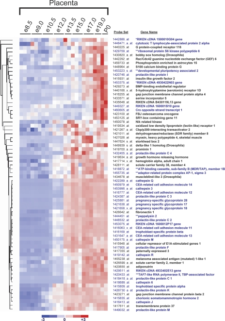
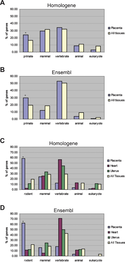
The invention of the placenta facilitated the evolution of mammals. How the placenta evolved from the simple structure observed in birds and reptiles into the complex organ that sustains human life is one of the great mysteries of evolution. By using a timecourse microarray analysis including the entire lifetime of the placenta, we uncover molecular and genomic changes that underlie placentation and find that two distinct evolutionary mechanisms were utilized during placental evolution in mice and human. Ancient genes involved in growth and metabolism were co-opted for use during early embryogenesis, likely enabling the accelerated development of extraembryonic tissues. Recently duplicated genes are utilized at later stages of placentation to meet the metabolic needs of a diverse range of pregnancy physiologies. Together, these mechanisms served to develop the specialized placenta, a novel structure that led to expansion of the eutherian mammal, including humankind.


Figures

Figure 1.
Figure 1.
Genome-wide expression profiling of the fetal and maternal placenta. (A) Samples were taken at nine stages throughout placental development, and dissected into placental and decidual portions. The data set includes biological triplicates for placenta samples at e8.5, e9.0, e10.5, e12.0, e15.0, and e17.0, biological duplicates for placenta samples at e13.5, e19.0, and P0, and biological duplicates for decidual samples at e8.5, e9.0, e10.5, e12.0, e15.0, e17.0, e19.0, and P0. (B) Linear correlation coefficients (r-values) between biological replicates range from 0.90 to 0.99 for fetally derived tissues and from 0.94 to 0.99 for maternally derived tissues, indicating a high degree of correlation between replicate samples at each timepoint. Microarray profiles for known placental hormones correspond with previously published expression patterns (C) and are confirmed by RT–PCR (D) (Linzer et al. 1985; Faria et al. 1991; Yotsumoto et al. 1998; Linzer and Fisher 1999; Soares 2004). Prl3d1 (chorionic somatomammotropin hormone 1); Prl3b1 (chorionic somatomammotropin hormone 2); Prl2c2 (proliferin); Adm (adrenomedullin) (represented by two probe sets). Figure 1A adapted by permission from Macmillan Publishers Ltd.: Nature Reviews Genetics (Rossant and Cross 2001), copyright 2001 (http://www.nature.com/nrg).
Figure 2.
Figure 2.
A molecular transition revealed at mid-gestation in the fetal placenta. The transition at mid-gestation is global, encompassing 3543 genes in the placenta and 1108 genes in the decidua that change significantly over time (FDR < 0.1% using SAM [Tusher et al. 2001] and having a minimum 1.5-fold change within a 2-d time period). Genes that change significantly during placental development were clustered using dCHIP (Li and Wong 2001a, b). Expression values for each gene across all samples were linearly scaled to have a mean of 0 and standard deviation of 1; red indicates expression greater than the mean; and blue indicates expression below the mean. (A) Clustergram of 3543 genes that change significantly in the placenta. (B) Clustergram of 1108 genes that change significantly in the decidua. Genes in Clusters labeled D are highly expressed in the developing placenta (e8.5–e13.5), while genes in Clusters labeled M are highly expressed in the mature placenta (e13.5–P0).
Figure 3.
Figure 3.
Placental genes have ancient origins. Evolutionary origins of preferentially expressed genes in the placenta (410 genes), decidua (347 genes), or in any tissue (3055 genes). The evolutionary origin of each gene was determined by finding the most distantly related species that has a one-to-one, reciprocal best-match ortholog identified in the Homologene database (A) or the Ensembl database (B). Asterisks (*) indicate significant enrichment over background (All Tissues). The placenta demonstrates significant enrichment for genes with eukaryotic orthology according to both Homologene (P < 1 × 10⁻¹⁹) and Ensembl (P < 1 × 10⁻²⁹). The decidua demonstrates significant enrichment for genes with vertebrate orthology according to both Homologene (P < 0.05) and Ensembl (P < 0.01).
Figure 4.
Figure 4.
Genes representing the mature phase reveal recent evolution by duplication and divergence. Evolutionary origins of placental preferentially expressed genes during the developing (329 genes) and mature (70 genes) phases of placentation. The evolutionary origin of each gene was determined by finding the most distantly related species that has a one-to-one, reciprocal best-match ortholog identified in the Homologene database (A) or the Ensembl database (B). Asterisks (*) indicate significant enrichment over background (All Tissues; 3055 genes). The developing placenta is significantly enriched for genes with eukaryotic orthology according to both Homologene (P < 1.0 × 10⁻²⁷) and Ensembl (P < 1.0 × 10⁻³⁵). In contrast, the mature placenta is significantly enriched for genes with rodent orthology according to both Homologene (P < 1.0 × 10⁻⁷) and Ensembl (P < 1.0 × 10⁻¹¹).
Figure 5.
Figure 5.
Newly evolved rodent-specific genes from the mature phase represent three distinct gene families. Among preferentially expressed genes, the mature cluster is highly enriched for genes that have evolved since rodents diverged from primates. Of the 37 rodent specific genes identified by either Homologene or Ensembl (see blue gene names), 29 are called rodent specific by both Homologene and Ensembl, two by Homologene only (*), and six by Ensembl only (**). Three large gene families account for a majority of the rodent-specific genes: the prolactin-like protein family, the CEA-related cellular adhesion molecule, and pregnancy-specific glycoprotein family, and the cathepsin family.
Figure 6.
Figure 6.
Primate-specific genes enriched in human mature placenta, indicating conservation of a gene duplication mechanism. Evolutionary origins of genes with PlacPEM ≥4 in mature placenta samples from the unscaled Novartis GeneAtlas human and mouse datasets using either Homologene (A) or Ensembl (B). Asterisks (*) indicate significant enrichment over background (All Tissues). In human, the GeneAtlas mature placenta gene set demonstrates significant enrichment of primate specific genes according to Homologene (P < 0.05) and Ensembl (P < 0.01). The GeneAtlas mature placenta gene set for the mouse is significantly enriched for rodent-specific genes according to both Homologene (P < 1.0 × 10⁻¹¹) (C) and Ensembl (P < 1.0 × 10⁻¹¹) (D). Genes with HeartPEM ≥ 4 (i.e., genes for which the heart expression level divided by the average expression is ≥4), UterusPEM ≥ 4, or PEM ≥ 4 in any tissue are included for comparison.

References

    1. Ain R., Dai G., Dunmore J.H., Godwin A.R., Soares M.J., Dai G., Dunmore J.H., Godwin A.R., Soares M.J., Dunmore J.H., Godwin A.R., Soares M.J., Godwin A.R., Soares M.J., Soares M.J. A prolactin family paralog regulates reproductive adaptations to a physiological stressor. Proc. Natl. Acad. Sci. 2004;101:16543–16548. - PMC - PubMed
    1. Alam S.M., Ain R., Konno T., Ho-Chen J.K., Soares M.J., Ain R., Konno T., Ho-Chen J.K., Soares M.J., Konno T., Ho-Chen J.K., Soares M.J., Ho-Chen J.K., Soares M.J., Soares M.J. The rat prolactin gene family locus: Species-specific gene family expansion. Mamm. Genome. 2006;17:858–877. - PubMed
    1. Beauchemin N., Draber P., Dveksler G., Gold P., Gray-Owen S., Grunert F., Hammarstrom S., Holmes K.V., Karlsson A., Kuroki M., Draber P., Dveksler G., Gold P., Gray-Owen S., Grunert F., Hammarstrom S., Holmes K.V., Karlsson A., Kuroki M., Dveksler G., Gold P., Gray-Owen S., Grunert F., Hammarstrom S., Holmes K.V., Karlsson A., Kuroki M., Gold P., Gray-Owen S., Grunert F., Hammarstrom S., Holmes K.V., Karlsson A., Kuroki M., Gray-Owen S., Grunert F., Hammarstrom S., Holmes K.V., Karlsson A., Kuroki M., Grunert F., Hammarstrom S., Holmes K.V., Karlsson A., Kuroki M., Hammarstrom S., Holmes K.V., Karlsson A., Kuroki M., Holmes K.V., Karlsson A., Kuroki M., Karlsson A., Kuroki M., Kuroki M., et al. Redefined nomenclature for members of the carcinoembryonic antigen family. Exp. Cell Res. 1999;252:243–249. - PubMed
    1. Carroll S.B. Evolution at two levels: On genes and form. PLoS Biol. 2005;3:e245. doi: 10.1371/journal.pbio.0030245. - DOI - PMC - PubMed
    1. Carter A.M., Mess A., Mess A. Evolution of the placenta in eutherian mammals. Placenta. 2006;28:259–262. - PubMed

Publication types