. 2008 Apr;118(4):1367-79.
doi: 10.1172/JCI33775.

The initial steps of ovarian cancer cell metastasis are mediated by MMP-2 cleavage of vitronectin and fibronectin

Affiliations

The initial steps of ovarian cancer cell metastasis are mediated by MMP-2 cleavage of vitronectin and fibronectin

Hilary A Kenny et al. J Clin Invest. 2008 Apr.

Abstract

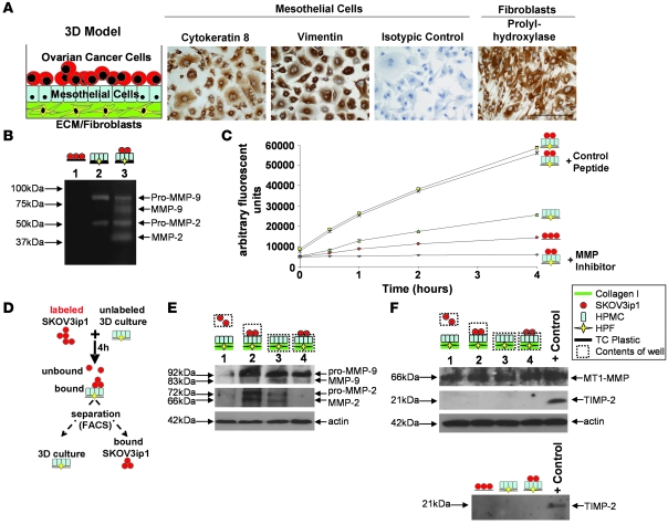
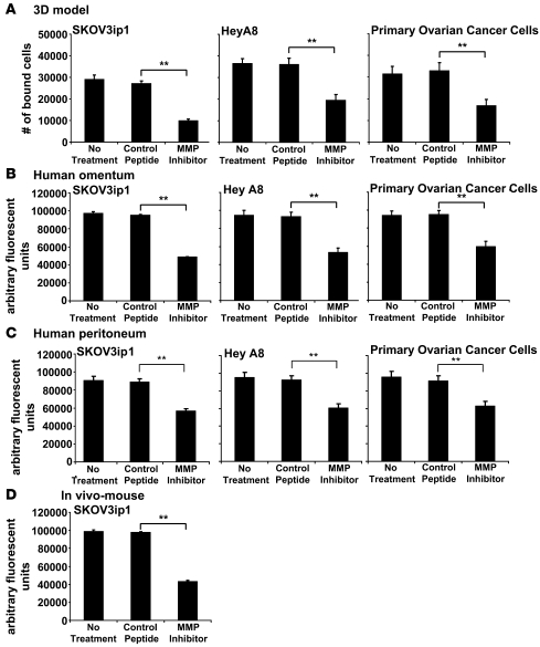
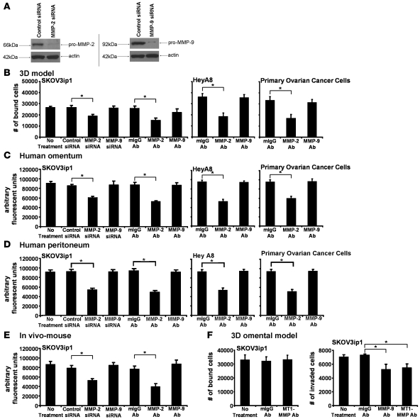
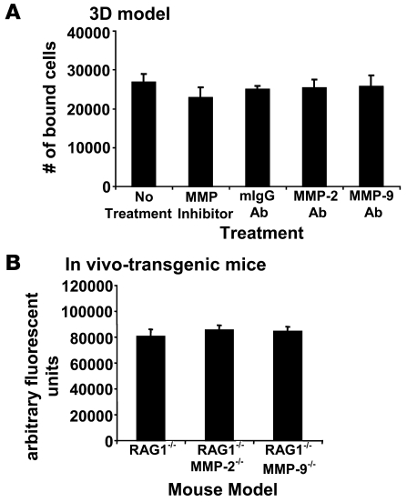
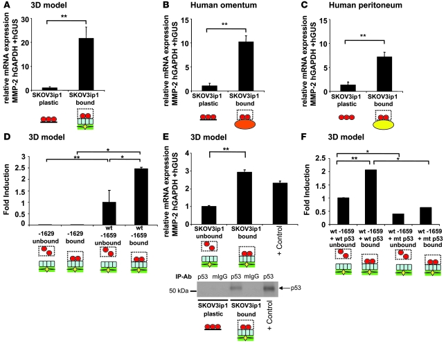
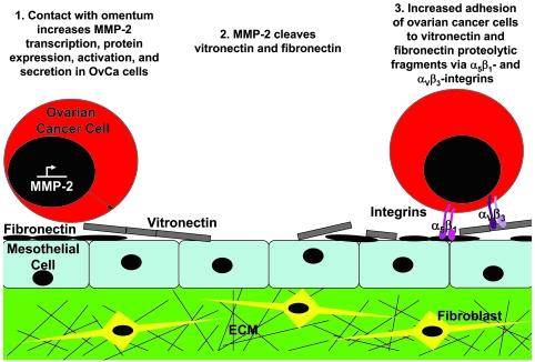
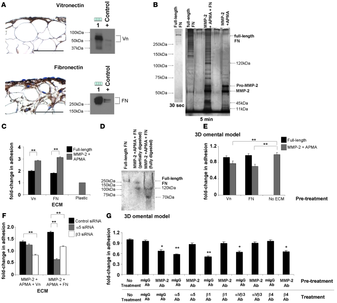
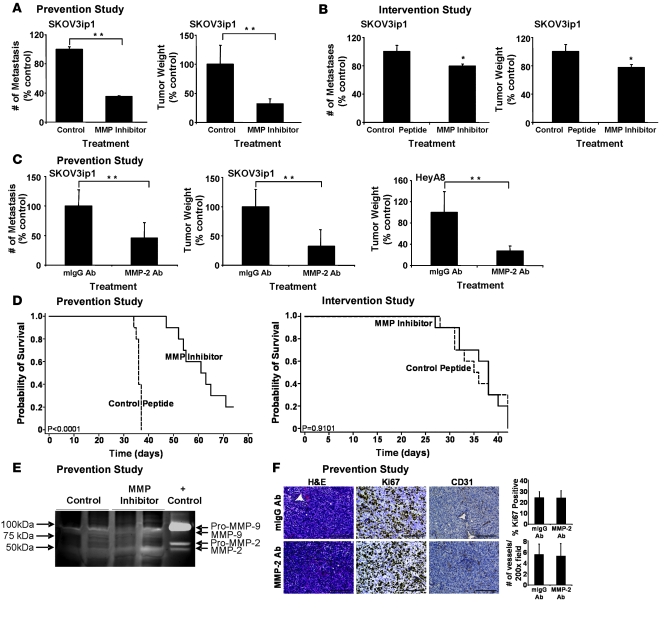
Most patients (80%) with ovarian cancer (OvCa) present with metastatic disease. Attachment of OvCa cells to peritoneum and omentum represents the first rate-limiting step for metastatic spread. Therefore, identifying factors regulating cell attachment in the abdominal cavity is critical to the development of therapeutic agents. We show here that MMP-2 expression was upregulated in OvCa cells upon attachment to their microenvironment. Downregulation of MMP-2 mRNA or pharmacological inhibition of MMP-2 proteolytic function, in both human OvCa primary cells and cell lines, reduced attachment of OvCa cells to a 3D organotypic model of metastatic OvCa, full human omentum or peritoneum, and in vivo to mouse peritoneum and omentum. Absence of MMP-2 in the host did not alter OvCa adhesion, as determined utilizing mice harboring homozygous null mutations in either the Mmp2 or Mmp9 genes. Conversely, adhesion induced upregulation of MMP-2 mRNA in OvCa cells. MMP-2 inhibition in OvCa cells through pharmacological or antibody treatment prior to i.p. dissemination in nude mice significantly decreased tumor growth and metastasis and extended survival. MMP-2 enhanced peritoneal adhesion of OvCa cells through cleavage of ECM proteins fibronectin (FN) and vitronectin (Vn) into small fragments and increased binding of OvCa cells to these FN and Vn fragments and their receptors, alpha5beta1 and alphaVbeta3 integrin. These findings indicate that MMP-2 expressed by metastatic OvCa cells functionally regulates their attachment to peritoneal surfaces.


Figures

Figure 1
Figure 1. Binding of OvCa cells to an omental 3D culture increases MMP-2/-9 expression and secretion in cancer cells.
(A) Concept for a 3D culture to imitate abdominal mesothelium. HPMCs stained with an antibody against cytokeratin 8, vimentin, or an isotypic specific control antibody. HPFs stained with a prolyl-hydroxylase antibody recognizing human fibroblasts. Scale bar: 50 μM. (B) Zymogram. Conditioned media from SKOV3ip1 cells (red circles), the 3D culture (composed of HPMCs [blue rectangles], HPFs [yellow diamonds], and collagen I [green rectangles]), or coculture was subjected to gelatin zymography. (C) Gelatinase assay. Cell-associated gelatinolytic activity in the indicated cell populations, with or without an MMP-2/-9 inhibitor, was determined using a quenched fluorogenic peptide. Fluorescence was measured with a fluorescence spectrophotometer. (D) Schematic of adhesion assay and subsequent cell sorting. Fluorescently labeled SKOV3ip1 cells were mixed with 3D culture for 4 h. The cells were mechanically removed from the plate and sorted by FACS. (E) Cell extracts were subjected to immunoblotting using an MMP-2 and MMP-9 specific antibody. The membrane was reprobed with an antibody against actin. Lane 1, unbound, floating SKOV3ip1 cells; lane 2, SKOV3ip1 cells that had attached to the 3D culture after FACS; Lane 3, 3D culture alone; Lane 4, 3D culture that had bound SKOV3ip1 cells after FACS. (F) Western blot for MT1-MMP and TIMP-2. SKOV3ip1 were plated on the 3D culture and then sorted as indicated in D. Cell extracts (top panel) or conditioned media (bottom panel) were subjected to immunoblotting with MT1-MMP or TIMP-2 antibody. HT-1080 CM was used as positive control.
Figure 2
Figure 2. An MMPI inhibits adhesion of OvCa cells to a 3D culture of human omentum, full human omentum and peritoneum, and mouse omentum.
SKOV3ip1, HeyA8, or primary human OvCa cells (50,000) were fluorescently labeled and added to the 3D culture (A), a piece of full human omentum (B), or full human peritoneum (C). The 3D culture, the human omentum, and the human peritoneum were washed with PBS, adherent cells on the omentum were lysed with NP-40, and fluorescence intensity was measured with a fluorescence spectrophotometer. (D) SKOV3ip1 cells were fluorescently labeled and 4 × 10⁶ were cells injected i.p. into nude mice (n = 3). The mice were sacrificed after 4 hours, omentum and peritoneum were lysed with NP-40, and fluorescence was measured. **P < 0.001. Each bar represents the mean of 3 wells and SD. Each graph is representative of at least 3 independent experiments.
Figure 3
Figure 3. Attachment of OvCa cells depends on MMP-2.
(A) SKOV3ip1 cells were transfected with a siRNA specific for MMP-2, MMP-9, or a control siRNA and plated on collagen type I, and MMP-2 and MMP-9 were detected by immunoblotting with specific antibodies. The membrane was reprobed with actin. (BD) Adhesion assays. SKOV3ip1 cells were transfected with a siRNA specific for MMP-2 and MMP-9. SKOV3ip1, HeyA8, and primary OvCa cells were pretreated with an MMP-2– or MMP-9–blocking antibody and then fluorescently labeled. OvCa cells were added to the 3D culture (B), a piece of full human omentum (C), or full human peritoneum (D), and the adherent cells quantified with a fluorescence reader as described in Figure 2. (E) SKOV3ip1 cells (1 × 10⁶) were labeled fluorescently and injected i.p. into nude mice for 4 hours (n = 3). Mice were sacrificed after 4 hours, omentum and peritoneum was lysed with NP-40, and fluorescence was measured. (F) Adhesion assay. SKOV3ip1 cells (5 × 10⁴) pretreated with the MT1-MMP antibody were added to the 3D culture and an adhesion assay performed as described in Figure 2 (left panel). Invasion assay (right panel). SKOV3ip1 cells (3 × 10⁴) were treated with a MT1-MMP– or MMP-9–blocking antibody, and invasion was analyzed after 24 hours. *P < 0.01.
Figure 4
Figure 4. Host-derived MMP-2 and MMP-9 does not affect adhesion of OvCa cells.
(A) The 3D culture was pretreated with either the cyclic peptide inhibiting preferentially MMP-2/-9 or a monoclonal antibody against MMP-9 or MMP-2, respectively. Subsequently, adhesion assay was performed as described in Figure 2. (B) Six-week-old RAG-1–/– mice, WT for MMP2 and MMP9 or RAG-1–/– either deficient in MMP2 (MMP2–/–) or MMP9 (MMP9–/–), were injected with fluorescently labeled SKOV3ip1. After 4 h, the mice were sacrificed, equal parts of omentum and peritoneum were lysed in NP-40, and fluorescence was measured.
Figure 5
Figure 5. MMP-2 is transcriptionally upregulated in OvCa cells upon interaction with host stroma.
SKOV3ip1 cells were added to the 3D culture (A), full human omentum (B), or full human peritoneum (C), and then cells were sorted by FACS. The relative expression of MMP-2 normalized to GAPDH, and huGUS was measured by TaqMan quantitative real-time RT-PCR. (D) SKOV3ip1 cells were transfected with 5 μg of the –1,659-bp (WT) or the –1,629-bp MMP-2 promoter and adhered to 3D model, and luciferase activity was measured. (E) The relative expression of p53 normalized to GAPDH, and huGUS was measured by TaqMan quantitative real-time RT-PCR (top panel). Protein lysates from SKOV3ip1 cells cultured on plastic or 3D culture (after sorting) were immunoprecipitated with control mouse IgG or monoclonal p53 antibody, and western blot analysis for p53 was conducted on lysates using a pantropic sheep anti-p53 antibody (bottom panel). Positive control was RKO mRNA or cell lysates. (F) SKOV3ip1 cells were cotransfected with 5 μg of the WT MMP-2 promoter and a WT or mutated p53 expression plasmid and adhered to the 3D model, and luciferase activity was measured. Luciferase activity was normalized to number of bound and unbound cells, and all assays were run in duplicate. *P < 0.01, **P < 0.001. Each graph is representative of 3 independent experiments.
Figure 8
Figure 8. Proposed role of MMP-2 in early OvCa cell metastasis to the omentum and peritoneum.
Figure 7
Figure 7. MMP-2 cleavage of Vn and FN increases OvCa adhesion.
(A) Human omentum and peritoneum stained with Vn- or FN-specific antibodies (left panel). HPMCs were subjected to immunoblotting (right panel) using Vn- and FN-specific antibodies. Lane 1, HPMCs. HT-1080 CM was used as positive control. Original magnification, ×400. (B) Full-length FN was incubated with activated MMP-2, and fragments were resolved on a 10% Tris-HCl gel by silver stain analysis. (C) Adhesion assay of SKOV3ip1 cells to full-length and MMP-2–cleaved Vn or FN coated plates as described in Figure 2. (D) FN and MMP-2–cleaved FN were run on a native gel and transferred to nitrocellulose. An adhesion assay was performed with 1.0 × 10⁷ SKOV3ip1 cells for 4 hours, the membrane was washed, fixed, and bound cells were stained. (E) Competition assays were conducted. SKOV3ip1 cells were preincubated with full-length Vn, MMP-2–cleaved Vn, full-length FN, or MMP-2–cleaved FN, and an adhesion assay to 3D coculture was conducted. (F) SKOV3ip1 cells were transfected with a siRNA specific for α5 or β3 integrin, and adhesion assay was performed to MMP-2–cleaved Vn or FN coated plates. (G) Fluorescently labeled SKOV3ip1 cells were pretreated with either an MMP-2 or a mouse isotype IgG antibody followed by treatment with α5, β1, αvβ3, β4 integrin, or mouse isotype IgG antibodies. Subsequently, an adhesion assay was performed on the 3D model. *P < 0.01, **P < 0.001. Each graph and picture is representative of 3 independent experiments. Scale bar: 100 μm.
Figure 6
Figure 6. Single pretreatment of OvCa Cells with an MMP-2/-9 inhibitor or an MMP-2 antibody inhibits peritoneal metastases and increases survival.
(A) Prevention study. SKOV3ip1 cells (1 × 10⁶) were pretreated with MMPI or cyclic peptide and injected i.p. into nude mice. After 28 days, the number of metastasis and tumor weight were determined. **P < 0.001. (B) Intervention study. SKOV3ip1 cells (1 × 10⁶) were injected i.p. into nude mice. After 14 days of tumor growth, mice were treated with the MMPI 3 times per week for 3 weeks. After 28 days mice were sacrificed, the number of metastasis and tumor weight were determined. The columns represent the mean and the bars the SD. *P < 0.01. (C) Prevention study. SKOV3ip1 cells or HeyA8 cells (1 × 10⁶) were pretreated with an MMP-2 antibody or the isotypic specific control antibody and then injected i.p. into nude mice. After 28 days mice were sacrificed, the number of metastasis and tumor weight were determined. **P < 0.001. (D) Survival in prevention and intervention studies. The treatment courses of the prevention and intervention studies were conducted as described above. Mice were sacrificed once they showed signs of distress, and Kaplan-Meier curves were calculated. (E) Tumor lysates from MMPI or control peptide treated mice were subjected to gelatin zymography as described in Figure 1B. (F) Sections of tumors from MMP-2 antibody or mIgG antibody–treated mice were stained with H&E, a Ki-67-specific antibody to detect proliferation, or a CD31-specific antibody to count microvessels. Scale bar: 100 μm.

References

    1. Cannistra S.A. Cancer of the ovary. N. Engl. J. Med. 2004;351:2519–2529. doi: 10.1056/NEJMra041842. - DOI - PubMed
    1. Doig T., Monaghan H. Sampling the omentum in ovarian neoplasia: when one block is enough. Int. J. Gynecol. Cancer. 2006;16:36–40. doi: 10.1111/j.1525-1438.2006.00273.x. - DOI - PubMed
    1. Egeblad M., Werb Z. Νew functions for the matrix metalloproteinases in cancer progression. Nat. Rev. Cancer. 2002;2:161–174. doi: 10.1038/nrc745. - DOI - PubMed
    1. Liotta L., Abe S., Robey P., Martin G. Preferential digestion of basement membrane collagen by an enzyme derived from a metastatic murine tumor. Proc. Natl. Acad. Sci. U. S. A. 1979;76:2268–2272. doi: 10.1073/pnas.76.5.2268. - DOI - PMC - PubMed
    1. Lakka S.S., et al. Synergistic down-regulation of urokinase plasminogen activator receptor and matrix metalloproteinase-9 in SNB19 glioblastoma cells efficiently inhibits glioma cell invasion, angiogenesis and tumor growth. Cancer Res. 2003;63:2454–2461. - PubMed

Publication types

MeSH terms