. 2008 Apr;118(4):1532-43.
doi: 10.1172/JCI33464.

Reversal of axonal loss and disability in a mouse model of progressive multiple sclerosis

Affiliations

Reversal of axonal loss and disability in a mouse model of progressive multiple sclerosis

Alexandre S Basso et al. J Clin Invest. 2008 Apr.

Abstract

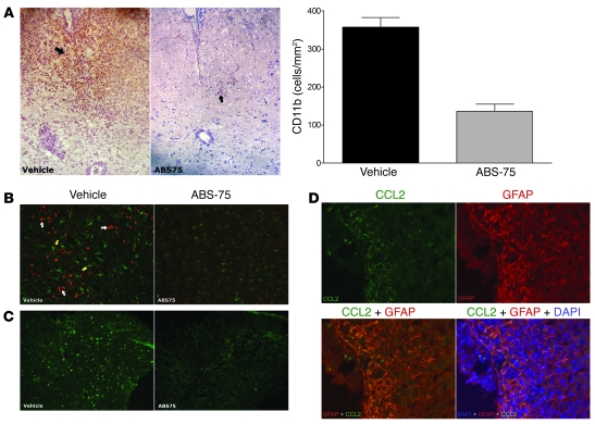
Axonal degeneration is an important determinant of progressive neurological disability in multiple sclerosis (MS). Thus, therapeutic approaches promoting neuroprotection could aid the treatment of progressive MS. Here, we used what we believe is a novel water-soluble fullerene derivative (ABS-75) attached to an NMDA receptor antagonist, which combines antioxidant and anti-excitotoxic properties, to block axonal damage and reduce disease progression in a chronic progressive EAE model. Fullerene ABS-75 treatment initiated after disease onset reduced the clinical progression of chronic EAE in NOD mice immunized with myelin-oligodendrocyte glycoprotein (MOG). Reduced disease progression in ABS-75-treated mice was associated with reduced axonal loss and demyelination in the spinal cord. Fullerene ABS-75 halted oxidative injury, CD11b+ infiltration, and CCL2 expression in the spinal cord of mice without interfering with antigen-specific T cell responses. In vitro, fullerene ABS-75 protected neurons from oxidative and glutamate-induced injury and restored glutamine synthetase and glutamate transporter expression in astrocytes under inflammatory insult. Glutamine synthetase expression was also increased in the white matter of fullerene ABS-75-treated animals. Our data demonstrate the neuroprotective effect of treatment with a fullerene compound combined with a NMDA receptor antagonist, which may be useful in the treatment of progressive MS and other neurodegenerative diseases.


Figures

Figure 1
Figure 1. Fullerene ABS-75 treatment reduces disease progression in secondary progressive EAE.
(A) Fullerene ABS-75 consists of the C60 fullerene core (i) attached to 4 adamantyl groups (ii) by oligoethyleneglycol bridges (iii). (B) Chronic progressive EAE was induced in 10-week-old NOD mice by subcutaneous immunization with 150 μg of MOG35–55 peptide in 4 mg/ml CFA. Pertussis toxin was given i.v. (150 ng per mouse) at the time of immunization and 48 h later. Fullerene ABS-75 (30 μg/kg, i.p.) was given daily beginning on day 20. n = 10 animals per group. Vehicle consisted of 2% DMSO. (C) Fullerene ABS-75 (30 μg/kg, i.p.) and memantine (1.5 mg/kg) were given daily beginning on day 19. n = 9–10 animals per group. Vehicle consisted of 2% DMSO. (D) Left: EAE was induced in 8-week-old SJL mice by s.c. immunization with 50 μg of PLP131–151 peptide in 4 mg/ml CFA. Pertussis toxin was given i.v. (150 ng per mouse) at the time of immunization and 48 h later. Fullerene ABS-75 (30 μg/kg, i.p.) and memantine (1.5 mg/kg) were given daily beginning on day 17. n = 15 animals per group. Arrows indicate relapses following the first attack. Right: Fullerene ABS-75 treatment (30 μg/kg, i.p.) begun 1 day before MOG immunization and done on a daily basis until the end of disease course was not able to ameliorate acute EAE in C57BL/6J mice. n = 8 animals per group. Disease score measured as follows: 0, no signs of disease; 1, loss of tone in the tail; 2, hindlimb paresis; 3, hindlimb paralysis; 4, tetraplegia; 5, moribund.
Figure 2
Figure 2. Fullerene ABS-75 treatment diminishes axonal loss and demyelination in secondary progressive EAE.
(A) Bielschowsky’s silver impregnation for axons showed a reduction of axonal density in the white matter of the spinal cord (arrows) of a vehicle-treated mouse as compared with ABS-75–treated animal. The axonal pathology analysis was performed after disease progression at the end of the disease course. Original magnification, ×50 (top panels); ×100 (bottom panels). Quantification of stained spinal cord sections by image analysis (ImageJ; NIH) confirmed a reduction in axonal loss when comparing ABS-75– and vehicle-treated animals. P < 0.02; Student’s t test, mean ± SD. (B) Luxol fast blue staining showed a reduction in the myelin content in the white matter of the spinal cord (arrows) of vehicle-treated mice as compared with ABS-75–treated animals. Original magnification, ×50 (top panels); ×100 (bottom panels). Quantification of stained spinal cord sections showed a reduction in demyelination when comparing ABS-75– and vehicle-treated animals. P < 0.01. Student’s t test, mean ± SD. (C) Neurofilament H staining, which identifies smaller nerve fibers (depicted in red; white arrows) shows reduction in nerve fiber density (percentage of the area within the ventral column of the white matter stained for neurofilament H) in controls (3.96% ± 1.43%) versus ABS-75 (16.13% ± 5.13%). P < 0.01; Student’s t test, mean ± SD. Nuclei of infiltrating cells stained blue (yellow arrows), and GFAP stained green. Original magnification, ×400. (D) Bielschowsky’s silver impregnation for axons showed no significant reduction of axonal density in the white matter of the spinal cord of SJL mice at the end of the disease course as compared with naive animals. Original magnification, ×100.
Figure 4
Figure 4. Fullerene ABS-75 protects neurons from oxidative injury and NMDA-dependent glutamate-induced injury but does not impair mouse memory function.
Rat cortical neurons (11 DIV) were challenged with either 60 μM H2O2 or 50 μM glutamate for 24 h in the absence or presence of ABS-75 (1 μM) or MK801 (1 μM). Glutamate- and H2O2-induced injuries were assessed by (A) MAP-2 staining and (B) LDH activity. The results showed reduced neuronal damage in fullerene ABS-75– and MK801-treated cultures. P < 0.01, control versus ABS-75 and MK801, and ABS-75 versus MK801. P < 0.01, control versus ABS-75, and control versus MK801 (H2O2-induced injury). ANOVA followed by post-hoc test, mean ± SD. (C) Fullerene ABS-75 and MK801 effects on memory function were assessed on a plus maze apparatus. On the first day (acquisition trial), animals were placed at the edge of an open arm and the latency to the first entry in the central area was recorded. On the second day (retention trial), all animals were returned to the arena and the latency to the first transfer onto the central area was recorded. Drugs were administered i.p. 30 min before the retention trial. All animals moved to the central area in the acquisition trial. In the retention trial, only animals treated with 0.15 mg/kg MK801 had similar results to those of vehicle mice in the acquisition trial. Animals treated with PBS, 2% DMSO, or ABS-75 arrived at the central area faster in the retention trial than in the acquisition trial, indicating that, unlike MK801, fullerene ABS-75 does not impair memory function. P < 0.05, Kruskal-Wallis test followed by Dunn’s multiple comparisons test. n = 10 animals per group. Median (line through each box) ± SD (error bars) are shown. Original magnification, ×130.
Figure 3
Figure 3. Fullerene ABS-75 treatment halts oxidative injury, blocks CCL2 production by astrocytes, and reduces CD11b infiltration into the spinal cord of secondary progressive EAE mice.
(A) CD11b staining revealed large numbers of CD11b+ cells infiltrating the ventral column (arrows) of vehicle-treated control animals, which was almost absent in mice that received the fullerene ABS-75 treatment. Original magnification, ×200. Quantification confirmed reduced CD11b+ cells infiltrating the spinal cord in ABS-75–treated animals with progressive EAE. P < 0.007; Student’s t test, mean ± SD. (B) Intense staining for nitrotyrosine (stained red; white arrows) in the spinal cord of vehicle-treated mice after disease progression indicated occurrence of oxidative injury, which was absent in ABS-75–treated animals. The presence of hyperreactive astrocytes (GFAP, stained green, yellow arrows) was associated with oxidative injury in vehicle-treated animals. Original magnification, ×200. (C) CCL2 staining demonstrated a large number of cells expressing CCL2 in the spinal cord of control animals after disease progression, whereas CCL2-positive cells were virtually absent in animals treated with fullerene ABS-75. Original magnification, ×100. (D) Triple staining performed on the spinal cord of control mice revealed high CCL2 expression (green) by astrocytic processes (GFAP, shown in red) in inflamed (blue nuclei of infiltrating cells) regions of the white matter. This demonstrates that in control animals, infiltration of inflammatory cells is associated with astrocytic expression of CCL2. Original magnification, ×400.
Figure 5
Figure 5. Fullerene ABS-75 rescues the reduced expression of EAAT1 and GS that occurs in astrocytes under inflammatory conditions.
(A) Primary mouse astrocytes were stimulated with either TNF-α (50 ng/ml) or LPS (1 μg/ml) and IFN-γ (200 U/ml) for 24 h in the absence or presence of ABS-75 (1 μM) or MK801 (1 μM). GS and EAAT1 expression were assessed by western blot. Quantification was performed using densitometry analysis (ImageJ; NIH) for 2 independent experiments. We found that both ABS-75 and MK801 could reverse the reduced expression of EAAT1 and GS by astrocytes under inflammatory conditions. P < 0.01, ANOVA followed by post-hoc test, mean ± SD. (B) Immunohistochemistry performed on spinal cord sections analyzed after disease progression revealed increased GS expression within the white matter of fullerene ABS-75 group as compared with vehicle-treated animals. Quantification by ImageJ confirmed increased numbers of GS-expressing glial cells in the white matter of fullerene ABS-75–treated mice. P < 0.05, Student’s t test, mean ± SD. (C) Immunohistochemistry performed on spinal cord sections analyzed after disease progression revealed that the cells expressing GS within the white matter were either oligodendrocytes (CNPase positive) or astrocytes (GFAP positive). Original magnification, ×200 (B); ×630 (C).
Figure 6
Figure 6. Fullerene ABS-75 treatment does not interfere with the adaptive compartment of the immune system.
(AC) Splenocytes from vehicle- and ABS-75–treated animals (4 per group) were harvested by the end of disease course and cultivated in the presence of MOG for (A) T cell proliferation, (B) IL-2, and (C) IFN-γ production. No differences were observed. (D) Serum collected from vehicle- and ABS-75–treated animals at the end of disease course was tested for MOG-specific IgG2b levels. No differences were observed. (E and F) NOD mice were treated once a day with fullerene ABS-75 (30 μg/kg i.p.) from day –1 to day 10 after immunization, when draining lymph nodes were harvested to evaluate T cell recall response upon MOG stimulation. No differences were found for (E) T cell proliferation and (F) IFN-γ production. (G and H) NOD mice were immunized with MOG, and after 10 days, cells harvested from the draining lymph nodes were stimulated with antigen in the presence or absence of 1 μM fullerene ABS-75. In vitro 1 μM fullerene ABS-75 did not interfere with (G) T cell proliferation and (H) IFN-γ production upon antigen-specific stimulation.

References

    1. Noseworthy J.H., Lucchinetti C., Rodriguez M., Weinshenker B.G. Multiple sclerosis. N. Engl. J. Med. 2000;343:938–952. - PubMed
    1. Sospedra M., Martin R. Immunology of multiple sclerosis. Annu. Rev. Immunol. 2005;23:683–747. doi: 10.1146/annurev.immunol.23.021704.115707. - DOI - PubMed
    1. Weinshenker B.G., et al. The natural history of multiple sclerosis: a geographically based study. I. Clinical course and disability. Brain. 1989;112:133–146. doi: 10.1093/brain/112.1.133. - DOI - PubMed
    1. Bjartmar C., Wujek J.R., Trapp B.D. Axonal loss in the pathology of MS: consequences for understanding the progressive phase of the disease. J. Neurol. Sci. 2003;206:165–171. doi: 10.1016/S0022-510X(02)00069-2. - DOI - PubMed
    1. Wujek J.R., et al. Axon loss in the spinal cord determines permanent neurological disability in an animal model of multiple sclerosis. J. Neuropathol. Exp. Neurol. 2002;61:23–32. - PubMed

Publication types

MeSH terms