

Ethanol selectively attenuates NMDAR-mediated synaptic transmission in the prefrontal cortex
- PMID: 18341645
- PMCID: PMC3587142
- DOI: 10.1111/j.1530-0277.2008.00625.x
Ethanol selectively attenuates NMDAR-mediated synaptic transmission in the prefrontal cortex
Abstract
Background: Brain imaging studies have revealed abnormal function in the prefrontal cortex (PFC) of alcoholics that may contribute to the impulsive behavior and lack of control over drinking that characterizes this disorder. Understanding how ethanol affects the physiology of PFC neurons may help explain this loss of control and lead to better treatments for alcohol addiction. In a previous study from this laboratory, we showed that ethanol inhibits complex patterns of persistent activity (known as "up-states") in medial PFC (mPFC) neurons in a reversible and concentration-dependent manner.
Methods: In the current study, whole-cell patch clamp recordings were used to directly examine the effects of ethanol on the glutamatergic and GABAergic components that underlie persistent activity.
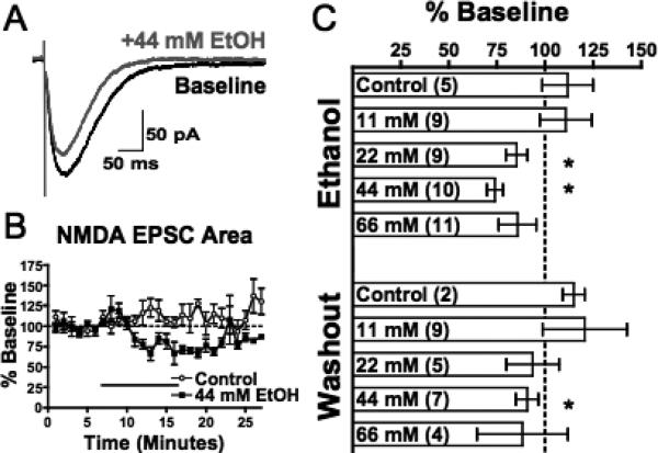
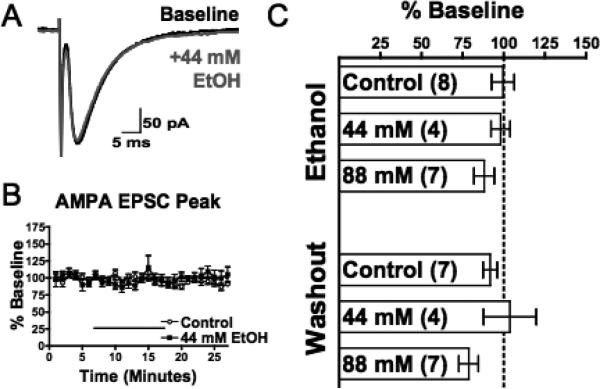
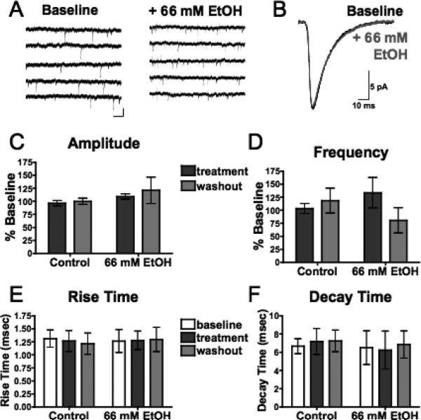
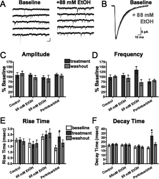
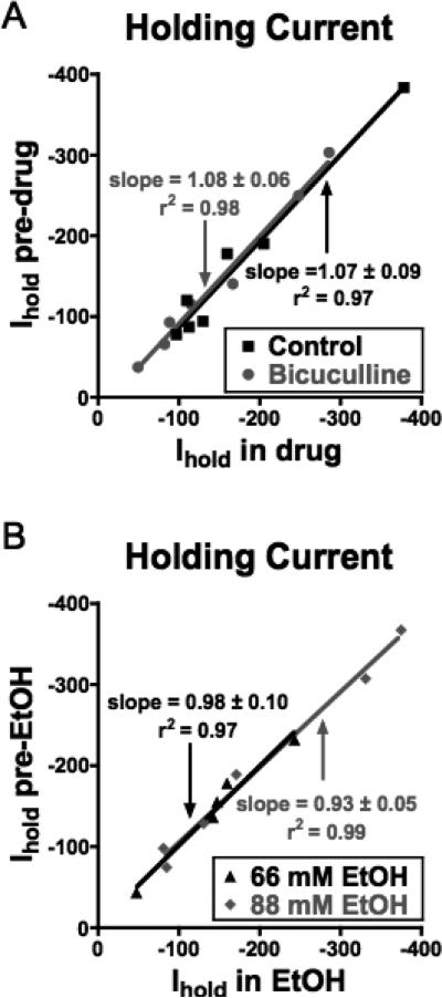
Results: In deep-layer mPFC pyramidal neurons, ethanol reversibly attenuated electrically evoked N-methyl-D-aspartate-type glutamate receptor (NMDAR)-mediated EPSCs. Significant inhibition was observed at concentrations as low as 22 mM, equivalent to a blood ethanol concentration (0.1%) typically associated with legal limits for intoxication. In contrast to NMDA responses, neither evoked nor spontaneous EPSCs mediated by alpha-amino-3-hydroxy-5-methylisoxazole-4-propionic acid-type glutamate receptor were affected by ethanol at concentrations as high as 88 mM, a concentration that can be fatal to non-tolerant individuals. At similar concentrations, ethanol also had little effect on spontaneous or evoked IPSCs mediated by a-type gamma-aminobutyric acid receptor. Finally, mPFC neurons showed little evidence of GABAR-mediated tonic current and this was unaffected by ethanol.
Conclusions: Together, these results suggest that NMDAR-mediated processes in the mPFC may be particularly susceptible to disruption following the acute ingestion of ethanol.
Figures

Similar articles
-
The NMDA-to-AMPA ratio at synapses onto layer 2/3 pyramidal neurons is conserved across prefrontal and visual cortices.J Neurophysiol. 2003 Aug;90(2):771-9. doi: 10.1152/jn.00070.2003. Epub 2003 Apr 2. J Neurophysiol. 2003. PMID: 12672778
-
Dopaminergic enhancement of excitatory synaptic transmission in layer II entorhinal neurons is dependent on D₁-like receptor-mediated signaling.Neuroscience. 2014 Jan 31;258:74-83. doi: 10.1016/j.neuroscience.2013.10.076. Epub 2013 Nov 9. Neuroscience. 2014. PMID: 24220689
-
Withdrawal from chronic alcohol impairs the serotonin-mediated modulation of GABAergic transmission in the infralimbic cortex in male rats.Neurobiol Dis. 2024 Sep;199:106590. doi: 10.1016/j.nbd.2024.106590. Epub 2024 Jul 10. Neurobiol Dis. 2024. PMID: 38996987
-
Understanding ethanol's acute effects on medial prefrontal cortex neural activity using state-space approaches.Neuropharmacology. 2021 Oct 15;198:108780. doi: 10.1016/j.neuropharm.2021.108780. Epub 2021 Sep 1. Neuropharmacology. 2021. PMID: 34480911 Free PMC article. Review.
-
Targeting prefrontal cortex GABAergic microcircuits for the treatment of alcohol use disorder.Front Synaptic Neurosci. 2022 Aug 29;14:936911. doi: 10.3389/fnsyn.2022.936911. eCollection 2022. Front Synaptic Neurosci. 2022. PMID: 36105666 Free PMC article. Review.
Cited by
-
Effects of acamprosate on attentional set-shifting and cellular function in the prefrontal cortex of chronic alcohol-exposed mice.Alcohol Clin Exp Res. 2015 Jun;39(6):953-61. doi: 10.1111/acer.12722. Epub 2015 Apr 23. Alcohol Clin Exp Res. 2015. PMID: 25903298 Free PMC article.
-
The abused inhalant toluene differentially modulates excitatory and inhibitory synaptic transmission in deep-layer neurons of the medial prefrontal cortex.Neuropsychopharmacology. 2011 Jun;36(7):1531-42. doi: 10.1038/npp.2011.38. Epub 2011 Mar 23. Neuropsychopharmacology. 2011. PMID: 21430649 Free PMC article.
-
Strychnine-sensitive glycine receptors on pyramidal neurons in layers II/III of the mouse prefrontal cortex are tonically activated.J Neurophysiol. 2014 Sep 1;112(5):1169-78. doi: 10.1152/jn.00714.2013. Epub 2014 May 28. J Neurophysiol. 2014. PMID: 24872538 Free PMC article.
-
Chronic Alcohol Exposure Induces Aberrant Mitochondrial Morphology and Inhibits Respiratory Capacity in the Medial Prefrontal Cortex of Mice.Front Neurosci. 2020 Oct 22;14:561173. doi: 10.3389/fnins.2020.561173. eCollection 2020. Front Neurosci. 2020. PMID: 33192248 Free PMC article.
-
Ethanol enhances glutamate transmission by retrograde dopamine signaling in a postsynaptic neuron/synaptic bouton preparation from the ventral tegmental area.Neuropsychopharmacology. 2009 Apr;34(5):1233-44. doi: 10.1038/npp.2008.143. Epub 2008 Sep 10. Neuropsychopharmacology. 2009. PMID: 18784647 Free PMC article.
References
-
- Borghese CM, Storustovu S, Ebert B, Herd MB, Belelli D, Lambert JJ, Marshall G, Wafford KA, Harris RA. The delta subunit of gamma-aminobutyric acid type A receptors does not confer sensitivity to low concentrations of ethanol. J Pharmacol Exp Ther. 2006;316:1360–1368. - PubMed
-
- Brickley SG, Revilla V, Cull-Candy SG, Wisden W, Farrant M. Adaptive regulation of neuronal excitability by a voltage-independent potassium conductance. Nature. 2001;409:88–92. - PubMed
-
- Chandra D, Jia F, Liang J, Peng Z, Suryanarayanan A, Werner DF, Spigelman I, Houser CR, Olsen RW, Harrison NL, Homanics GE. GABAA receptor alpha 4 subunits mediate extrasynaptic inhibition in thalamus and dentate gyrus and the action of gaboxadol. Proc Natl Acad Sci U S A. 2006;103:15230–15235. - PMC - PubMed
Publication types
MeSH terms
Substances
Grants and funding
LinkOut - more resources
Full Text Sources
Miscellaneous
