

Prostate mechanical imaging: a new method for prostate assessment
- PMID: 18342178
- PMCID: PMC2323601
- DOI: 10.1016/j.urology.2007.11.021
Prostate mechanical imaging: a new method for prostate assessment
Abstract
Objectives: To evaluate the ability of prostate mechanical imaging (PMI) technology to provide an objective and reproducible image and to assess the prostate nodularity.
Methods: We evaluated the PMI device developed by Artann Laboratories in a pilot clinical study. For the 168 patients (ages 44 to 94) who presented to an urologist for prostate evaluation, PMI-produced images and assessment of prostate size, shape, consistency/hardness, mobility, and nodularity were compared with digital rectal examination (DRE) findings. The PMI and DRE results were further tested for correlation against a transrectal ultrasound of the prostate (TRUS) guided biopsy for a subgroup of 21 patients with an elevated prostate-specific antigen level.
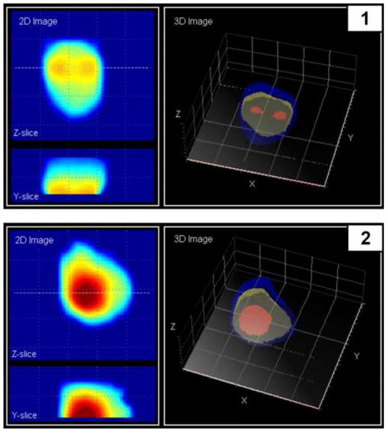
Results: In 84% of the cases, the PMI device was able to reconstruct three-dimensional (3D) and 2D cross-sectional images of the prostate. The PMI System and DRE pretests were able to determine malignant nodules in 10 and 6 patients, respectively, of the 13 patients with biopsy-confirmed malignant inclusions. The PMI System findings were consistent with all 8 biopsy negative cases, whereas the DRE had 1 abnormal reading for this group. The correlation between PMI and DRE detection of palpable nodularity was 81%, as indicated by the area under the receiver operating characteristic curve. Estimates of the prostate size provided by PMI and DRE were statistically significantly correlated.
Conclusions: The PMI has the potential to enable a physician to obtain, examine, and store a 3D image of the prostate based on mechanical and geometrical characteristics of the gland and its internal structures.
Figures

References
-
- Stephenson RA. Prostate cancer trends in the era of prostate-specific antigen. An update of incidence, mortality, and clinical factors from the SEER database. Urol Clin North Am. 2002;29:173–81. - PubMed
-
- Ilic D, O'Connor D, Green S, et al. Screening for prostate cancer. Cochrane Database Syst Rev. 2006;3:CD004720. - PubMed
-
- Gohagan JK, Prorok PC, Hayes RB, et al. The Prostate, Lung, Colorectal and Ovarian (PLCO) Cancer Screening Trial of the National Cancer Institute: history, organization, and status. Control Clin Trials. 2000;21:251S–272S. - PubMed
-
- Schroder FH. The European Screening Study for Prostate Cancer. Can J Oncol. 1994;4 1:102–5. discussion 106-9. - PubMed
-
- Draisma G, Boer R, Otto SJ, et al. Lead times and overdetection due to prostate-specific antigen screening: estimates from the European Randomized Study of Screening for Prostate Cancer. J Natl Cancer Inst. 2003;95:868–78. - PubMed
Publication types
MeSH terms
Grants and funding
LinkOut - more resources
Full Text Sources
Medical
