. 2008 May 1;473(1):42-7.
doi: 10.1016/j.abb.2008.02.036. Epub 2008 Feb 29.

Function and structure of lipid storage droplet protein 1 studied in lipoprotein complexes

Affiliations

Function and structure of lipid storage droplet protein 1 studied in lipoprotein complexes

Estela L Arrese et al. Arch Biochem Biophys. .

Abstract

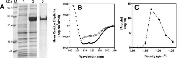
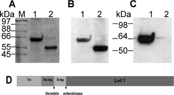
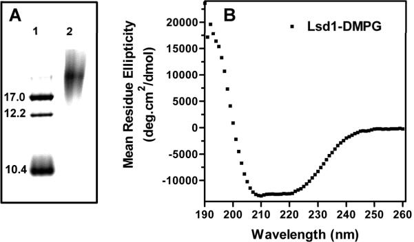
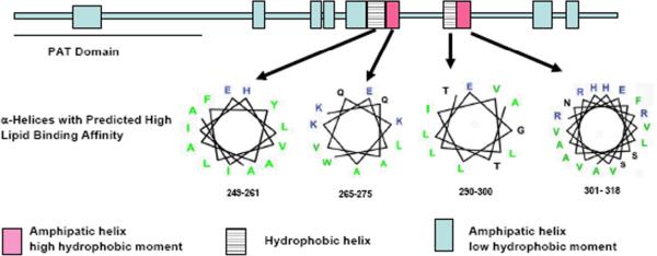
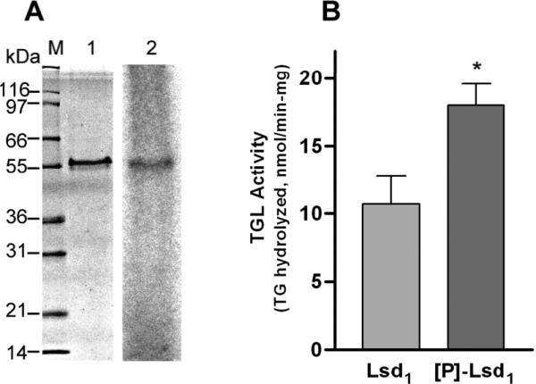
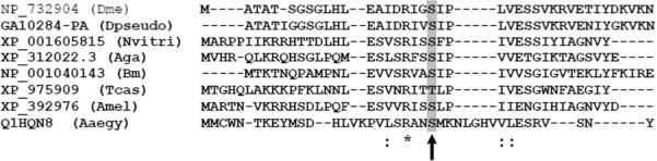
Triglycerides (TG) stored in lipid droplets (LDs) are the main energy reserve in all animals. The mechanism by which animals mobilize TG is complex and not fully understood. Several proteins surrounding the LDs have been implicated in TG homeostasis such as mammalian perilipin A and insect lipid storage proteins (Lsd). Most of the knowledge on LD-associated proteins comes from studies using cells or LDs leaving biochemical properties of these proteins uncharacterized. Here we describe the purification of recombinant Lsd1 and its reconstitution with lipids to form lipoprotein complexes suitable for functional and structural studies. Lsd1 in the lipid bound state is a predominately alpha-helical protein. Using lipoprotein complexes containing triolein it is shown that PKA mediated phosphorylation of Lsd1 promoted a 1.7-fold activation of the main fat body lipase demonstrating the direct link between Lsd1 phosphorylation and activation of lipolysis. Serine 20 was identified as the Lsd1-phosphorylation site triggering this effect.


Figures

Figure 1
Figure 1. Characterization of Trx-Lsd1
A) SDS-PAGE: lane 1, bacterial lysate; lane2, bacteria lysate after IPTG; lane 3, purified Trx-Lsd1. B) Far-UV CD spectrum of Trx-Lsd1 in 2M urea buffer (□), and Trx-Lsd1/DMPG in phosphate buffer (●). C) Flotation density of Trx-Lsd1/DMPG in a KBr density gradient.
Figure 2
Figure 2. Proteolytic Cleavage of Trx-Lsd1 in DMPG Complexes
A) SDS-PAGE: Samples of Trx-Lsd1 uncut (lane 1), and after thrombin cleavage (lane 2) were separated by electrophoresis. B-C) Western blot analysis of the samples shown in panel A using anti S-tag (B) and anti His (C) antibodies.
Figure 3
Figure 3. Secondary structure of rLsd1
A) Nondenaturing gel electrophoresis of rLsd1/DMPG complexes: Markers (Stoke diameters in nm) (lane 1); Lsd1/DMPG complexes (lane 2). B) CD spectrum of rLsd1 bound to DMPG.
Figure 4
Figure 4. Predicted α-helical regions of Lsd1
Helical regions of Lsd1 (431aa) are indicated with rectangles. The PAT domain is located between residues 17–170. The locations of the 11 predicted helices are: 44–63, 170–178, 211–219, 221–228, 236–247, 249–261, 265–275, 290–300, 301–318, 357–371, and 380–389. Helical wheel diagrams of the four helices having a high lipid binding affinity are shown (charged residues are in blue, hydrophobic residues in green and uncharged polar residues in black).
Figure 5
Figure 5. Effect of Lsd1 phosphorylation on TGL activity
A) Lsd1 bound to DMPG was phosphorylated by PKA and [γ-32P]-ATP followed by SDS-PAGE (1) and autoradiography (2). B) TGL activity was assayed against rLsd1/DMPG/[3H]-triolein containing unphosphorylated (Lsd1) or phosphorylated ([P]-Lsd1) protein. Data represent the mean ± SEM (n=10). Statistical comparisons were made by Student's t test. Two tail P value was significant (*P=0.012).
Figure 6
Figure 6. Alignment of the N-terminal regions of Lsd1
The alignment of the eight available Lsd1 sequences was performed using the T-Coffee program available at www.ebi.ac.uk/t-coffee. The accession numbers for the sequences are displayed in the figure. Dme, Drosophila melanogaster; Dpseudo, Drosophila pseudoobscura; Nvitri, Nasonia vitripennis; Aga, Anopheles gambiae; Bm, Bombyx mori; Tcas, Tribolium castaneum; Ame, Apis mellifera; Aaegy, Aedes aegypti. The arrow indicates Ser20. In Lsd1 from Tribolium castaneum Ser 20 is replaced by a threonine residue.

References

    1. Wolins NE, Brasaemle DL, Bickel PE. A proposed model of fat packaging by exchangeable lipid droplet proteins. FEBS Lett. 2006;580:5484–5491. - PubMed
    1. Ziegler R, Van Antwerpen R. Lipid uptake by insect oocytes. Insect Biochem Mol Biol. 2006;36:264–272. - PubMed
    1. Beenakkers AM, Van der Horst DJ, Van Marrewijk WJ. Insect lipids and lipoproteins, and their role in physiological processes. Prog Lipid Res. 1985;24:19–67. - PubMed
    1. Brasaemle DL. The perilipin family of structural lipid droplet proteins: Stabilization of lipid droplets and control of lipolysis. J Lipid Res. 2007 - PubMed
    1. Murphy DJ. The biogenesis and functions of lipid bodies in animals, plants and microorganisms. Prog Lipid Res. 2001;40:325–438. - PubMed

Publication types

MeSH terms