. 2008 Apr 15;316(2):359-70.
doi: 10.1016/j.ydbio.2008.01.042. Epub 2008 Feb 13.

Sox8 is a critical regulator of adult Sertoli cell function and male fertility

Affiliations

Sox8 is a critical regulator of adult Sertoli cell function and male fertility

Moira K O'Bryan et al. Dev Biol. .

Abstract

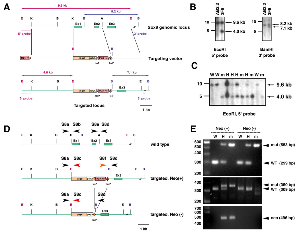
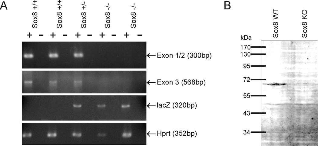
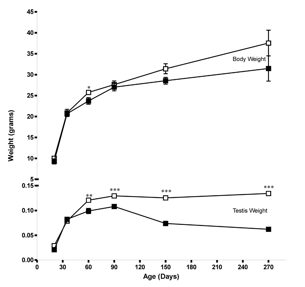
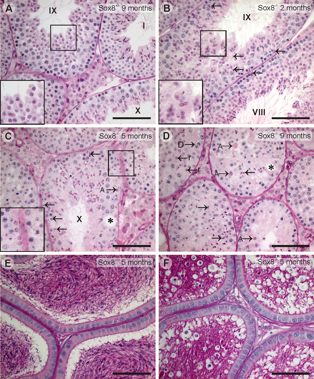
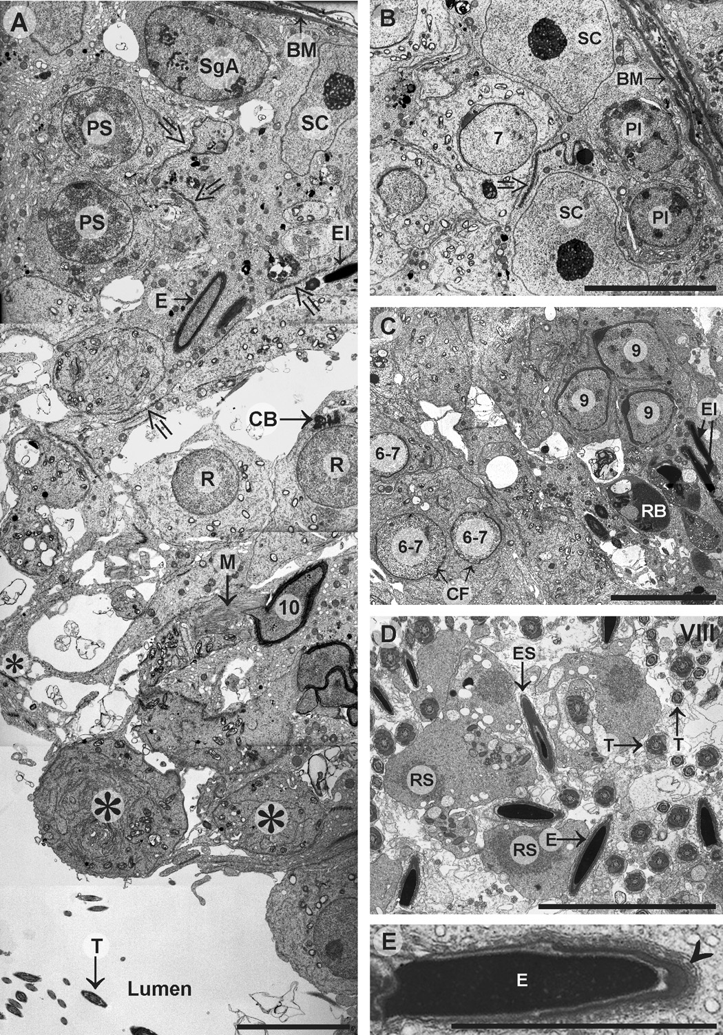
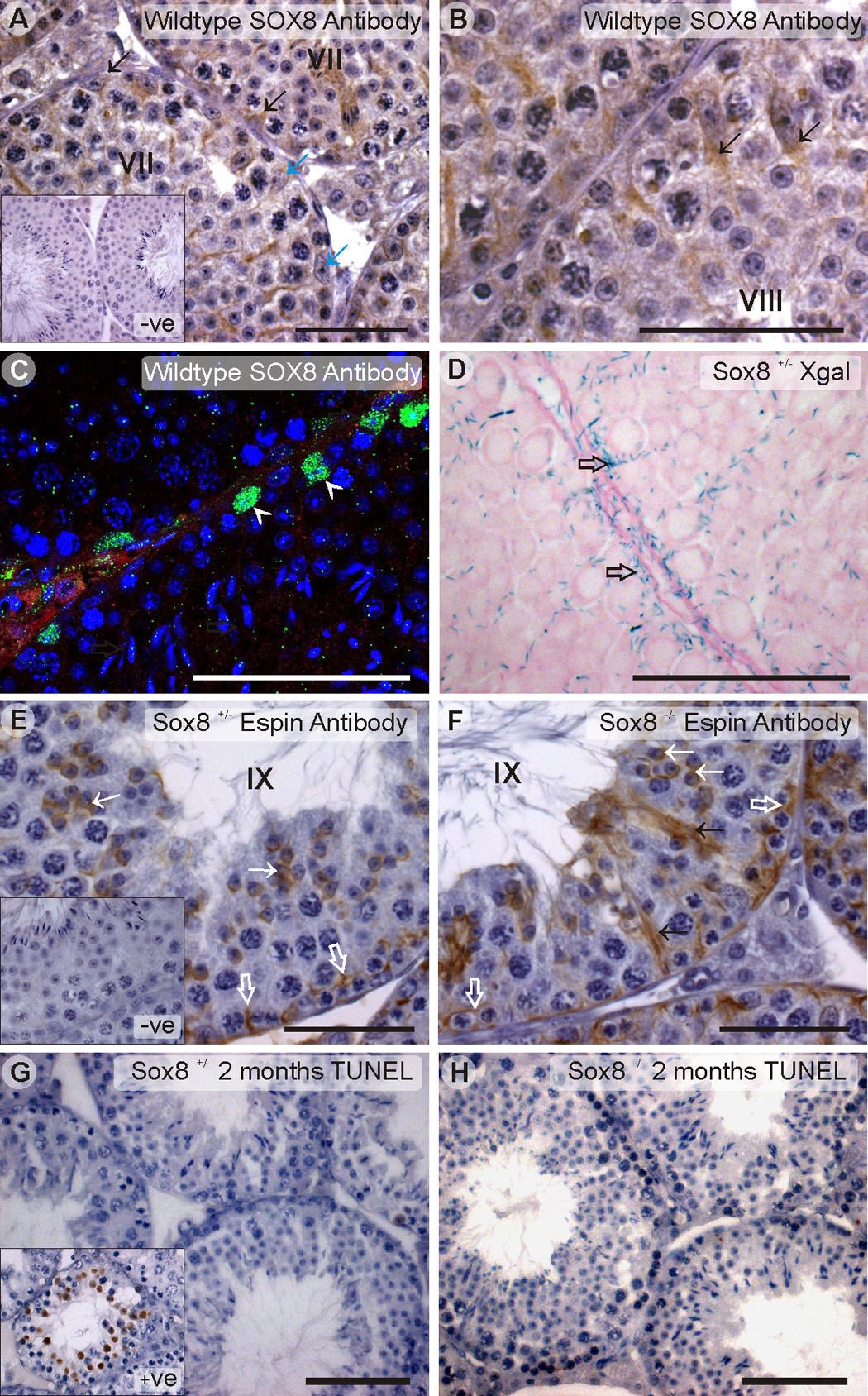
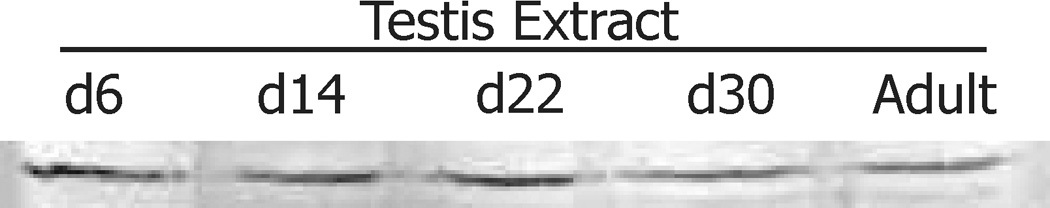
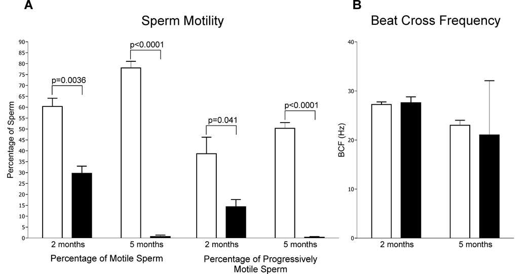
Sox8 encodes a high-mobility group transcription factor that is widely expressed during development. Sox8, -9 and -10 form group E of the Sox gene family which has been implicated in several human developmental disorders. In contrast to other SoxE genes, the role of Sox8 is unclear and Sox8 mouse mutants reportedly showed only idiopathic weight loss and reduced bone density. The careful analysis of our Sox8 null mice, however, revealed a progressive male infertility phenotype. Sox8 null males only sporadically produced litters of reduced size at young ages. We have shown that SOX8 protein is a product of adult Sertoli cells and its elimination results in an age-dependent deregulation of spermatogenesis, characterized by sloughing of spermatocytes and round spermatids, spermiation failure and a progressive disorganization of the spermatogenic cycle, which resulted in the inappropriate placement and juxtaposition of germ cell types within the epithelium. Those sperm that did enter the epididymides displayed abnormal motility. These data show that SOX8 is a critical regulator of adult Sertoli cell function and is required for both its cytoarchitectural and paracrine interactions with germ cells.


Figures

Figure 1
Figure 1
Generation of lacZ knock in mouse for Sox8. A. Targeting construct. An XbaI-AccI 2.6kb fragment containing exons 1 and 2 was replaced by a lacZ expression cassette followed by a floxed Pgk-neo cassette. Positions of 5’ and 3’ external probes for Southern analyses and expected band sizes are shown. A, AccI; B, BamHI; RI, EcoRI, K, KpnI; X, XbaI. B. Confirmation of targeting event in ES cells. Genomic DNA from wild-type ES cells (AS2.2) and correctly targeted cells (clone 3F9) were digested with an enzyme and probed as indicated. C. Interbreeding of heterozygous Sox8 mutant mice generated homozygous mutant mice. All pups from one litter were genotyped at the stage of weaning. D. Schematic representation of structure of each allele and positions of genotyping primers. Primers S8a, S8b, and S8c were used to differentiate the lacZ knock-in allele from WT, whereas primers S8d, S8e and S8f were used to differentiate the lacZ knock-in allele from the lacZ knock-in allele without the neo cassette. E. Examples of genotyping results. Combination of S8a, S8b and S8c differentiated all three genotype regardless the presence of the neo cassette (top). Combination of S8d, S8e, and S8f differentiated the lacZ knock-in allele with the neo cassette from the lacZ knock-in allele without the cassette (middle). Removal of the neo cassette was confirmed with primers that amplified inside of the cassette (bottom). W, wild type; H, heterozygous mutant; m, homozygous mutant. (Sox8 wildtype) Sox8−/− (knockout).
Figure 2
Figure 2
A. An assessment of Sox8 mRNA production in wild-type (WT), heterozygous (Het) and knockout (mut) mice. Testicular mRNA was extracted from 10 week old animals and analyzed by RT-PCR to assay for cDNA derived from exon 1–2 (targeted exons), cDNA derived from exon 3 which remained in situ and LacZ expression (which was targeted into the Sox8 locus). mRNA quality was assessed using Hprt expression. Product size is indicated in the brackets and RT (+) and no RT (−) is marked. B. The specificity of the anti-SOX8 serum as determined by Western blotting of adult testis extract from Sox8+/+ and Sox8−/− animals, showing SOX8 expression in the Sox8+/+ but not Sox8−/− animals.
Figure 3
Figure 3
Sox8−/− animals show an age-dependent effect on testis weight which is not dependent on a reduction in body weight. Testis and body weights were recorded for 4–26 animals per group and plotted +/− SEM. Age in days is indicated along the X-axis. *p<0.05, **p<0.01, ***p<0.001. Open squares, Sox8+/−,+/− (Sox8 heterozygotes), closed squares, Sox8−/− (Sox8 knockouts).
Figure 4
Figure 4
Testis and epididymal histology of Sox8+/− and (Sox8 heterozygotes) and Sox8−/− (Sox8−/− knockout) animals. A minimum of 3 mice of each age and genotype were examined. A. A testis section from a 9-month-old Sox8+/− animal showing normal seminiferous tubule histology. The inset illustrates the correct orientation of elongating spermatids in a stage IX tubule ie. with the acrosomes pointing towards the basement membrane. B. A 2-month-old Sox8−/− testis showing the presence of all germ cell types, but an increased abundance of retained elongated spermatids at stage IX (arrows) ie. spermiation failure. The inset shows the relative disorientation of elongating spermatids at stage IX. Spermiation failure and spermatid disorientation were also visible in stage VIII tubules. C A 5-month-old Sox8−/− testis showing a relative reduction in elongating spermatid numbers compared to control animals, increased spermiation failure (arrows) and increased epithelial vacuolation (asterisks) indicative of recent germ cell loss. The inset shows the inappropriate placement of step ~8 round spermatids (normally stage VIII) near the basement membrane of a largely stage X tubule. D. A 9-month- old Sox8−/− testis showing massive disorganization of the seminiferous epithelium with the presence of elongated spermatids in virtually every tubule, loss of round germ cells, the suggestion of increased apoptosis (A) and the inappropriate placement and timing / juxtaposition of germ cells within the epithelium. E. A section of epididymis from a 5-month-old Sox8+/− animal showing normal epididymal histology and an abundance of sperm within the lumen. F. A section of epididymis from a 5-month-old Sox8−/− animal showing the presence of normal epididymal epithelium, but a relative absence of sperm and a massive increase in the number of round germ cells within the lumen. The interstitial tissue of Sox8−/− animals appeared normal at all ages. The age and genotype of animals are indicated in the top right hand corner. The stage of the seminiferous epithelium cycle is indicated in roman numerals. The scale bars represent 100µm. Abbreviations: A – apoptotic cells; As – type A spermatogonia. Asterisk – vacuoles. Arrow – retained elongated spermatids (spermiation failure).
Figure 5
Figure 5
Electron microscopic examination of Sox8−/− (Sox8 knockout) testes. 2 mice of each age were examined. Panels A–C: 5 month old animals; panel D – a 2 month old animal. A. Even with advancing age, all stages of sperm development were visible within the seminiferous epithelium, however, as animals aged the placement of germ cells and the synchronization of germ cells development (the cycle of the seminiferous epithelium) became increasingly disorganized. For example, early stage round spermatids (R, step 1–5 as indicated by the lack of acrosome development) were found in the same tubule as elongating (step 10) spermatids and elongating spermatids. The epithelium contained many vacuoles and the Sertoli cell cytoplasm appeared lacey (*), indicative of recent germ cell loss. Inter-Sertoli cell junctions (the blood-testis barrier, large open arrows) appeared normal. B. Shows the inappropriate placement of step 7 round spermatids close to a Sertoli cell nucleus and the blood-testis barrier (open arrows). Sertoli cell nuclei appeared normal. C. Illustrates the deregulation of the seminiferous cycle whereby a cluster of step 6–7 round spermatids were seen in the same tubule as a cluster of step 9 elongating spermatids. There was also evidence of failed cytokinesis (CF), or the collapse of cytoplasmic bridges, between sister spermatids. The elongated spermatids were likely the result of a failure of spermiation at step 16 (stage VIII). D. A stage VIII tubule from a two month old animal showing a failure of ectoplasmic specialization (ES) resolution which usually occurs at stage VII. Sperm tail cross-sections appeared normal, consistent with the maintenance of sperm motility at least in some sperm. E. A retained spermatid showing the presence of actin bundles (the defining feature of the apical ectoplasmic specializations; arrowhead). Abbreviations: BM – basement membrane; CB – chromatoid body; CF – cytokinesis failure; E – elongating spermatid; El – elongated spermatid; M – manchette; PS – pachytene spermatocyte; Pl – preleptotene spermatocyte; R – round spermatid; RB – residual body; RS – aggregations of ribosomes; SC –Sertoli cell; SgA – type A spermatogonia; T – sperm tail. Large open arrows – inter-Sertoli cell junctions / the blood-testis-barrier, Arrowhead- actin bundles. Roman numerals – the stage of spermatogenesis. Arabic numbers – the step of spermiogenesis. Scale bars = 10 µm (A–D) 5µm (E).
Figure 6
Figure 6
A. and B. Immunohistochemical localization of SOX8 in wild-type testes showing Sertoli cells which were positive for both nuclear and cytoplasmic staining (black arrows) and other which contained only cytoplasmic staining (blue arrows). The inset in panel A shows negative control staining. C. Immunofluorescent localization of SOX8 (green) in wild-type testes showing nuclear Sertoli cell staining (white arrowheads). Red staining indicates E-cadherin and blue is DAPI. D. LacZ staining of a Sox8+/− testis showing the localization of SOX8 specifically to Sertoli cells. Open arrows indicate Sertoli cell cytoplasm. Germ cells showed no evidence of LacZ staining. E. and. F. The distribution of ectoplasmic specializations, as indicated by Espin immunolocalization, in 2-month-old Sox8+/− (E) and Sox8−/− (F) animals. White open arrows indicate the expression of the Sertoli cells derived blood-testis barrier at a level approximately 1 cells depth above the basement membrane ie. separating the spermatogonia and leptotene spermatocytes (basal compartment) from the meiotic and haploid (luminal) compartment. Staining for Espin was also observed around the apical aspect of spermatids from step 8 of spermatid development onwards (white arrows, indicative of the apical ectoplasmic specialization). Black arrows indicate Espin staining on retained elongated spermatids (spermiation failure). The inset on panel E shows a negative control section. G and H. TUNEL staining to detect apoptotic cells in 2 month old Sox8+/− (G) and Sox8−/− (H) animals. The inset on panel G shows a positive control section. +ve – positive control (wildtype), −ve – negative control (no antibody). Scale bars A-F= 50 µm, G and H =100 µm. Roman numerals indicate the stage of spermatogenesis. >3 animals were stained and examined.
Figure 7
Figure 7
The production of SOX8 in the post-natal testis. SOX8 was seen at all ages examined, but relative levels decreased with age consistent with expression by Sertoli cells.
Figure 8
Figure 8
Motility parameters of caudal epididymal sperm from 2- and 5-month-old Sox8+/− (Sox8 heterozygotes) (open bars) and Sox8−/− (Sox8 knockout) males (closed bars). A. Sox8−/− males showed an age-dependent reduction in the percentage of motility sperm produced, and the percentage of progressive motile sperm produced. B. In comparison, the beat cross frequency (the frequency with which a sperm track crosses the cell path in either direction), did not change significantly with age.

Similar articles

Cited by

References

    1. Beardsley A, O'Donnell L. Characterization of normal spermiation and spermiation failure induced by hormone suppression in adult rats. Biol. Reprod. 2003;68:1299–1307. - PubMed
    1. Bondurand N, et al. A molecular analysis of the yemenite deaf-blind hypopigmentation syndrome: SOX10 dysfunction causes different neurocristopathies. Hum. Mol. Genet. 1999;8:1785–1789. - PubMed
    1. Bowles J, et al. Phylogeny of the SOX family of developmental transcription factors based on sequence and structural indicators. Dev. Biol. 2000;227:239–255. - PubMed
    1. Cameron D, Griffin F. Ultrastructure of Sertoli-germ cell interactions in the normal and pathologic testis. In: M-GF R, editor. Male Reproduction: A multidisciplinary overview. Spain: Churchill; 1998. pp. 229–242.
    1. Chaboissier MC, et al. Functional analysis of Sox8 and Sox9 during sex determination in the mouse. Development. 2004;131:1891–1901. - PubMed

Publication types

MeSH terms