Review
. 2008 May;20(2):221-35, vi-vii.
doi: 10.1016/j.coms.2007.12.008.

Neurovascular pains: implications of migraine for the oral and maxillofacial surgeon

Affiliations
Review

Neurovascular pains: implications of migraine for the oral and maxillofacial surgeon

Donald R Nixdorf et al. Oral Maxillofac Surg Clin North Am. 2008 May.

Abstract

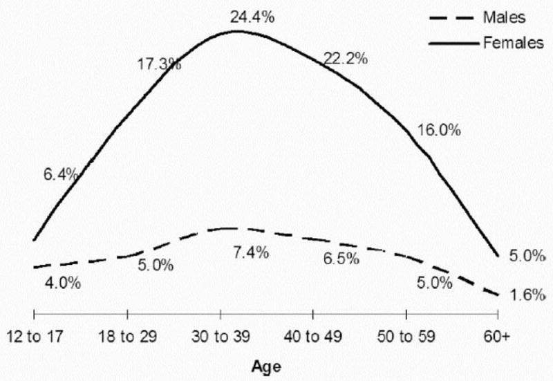
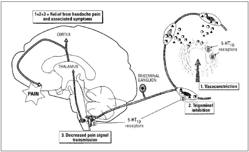
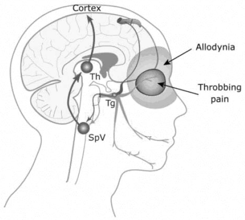
Epidemiologic studies have shown that migraine headaches are a common finding in the general population, often associated with a high degree of disability. Additionally, migraine has a reported comorbidity with other medical conditions, most notably with chronic pains, such as temporomandibular disorders. The pathophysiologic mechanisms involved with migraine are suggestive of an increased and prolonged hyperexcitability to stimuli, especially within the trigeminal distribution. Because migraine is mediated by branches of the trigeminal nerve it has the potential to mimic other types of pains, such as toothache or sinusitis. It is therefore recommended that oral and maxillofacial surgeons be familiar with the diagnostic criteria for migraine headaches to identify and appropriately treat such individuals who present to their clinics.


Figures

Figure 1
Figure 1. Prevalence of Migraine Headache Separated by Gender
The presence of migraine is considerably higher in females, especially during the ages of 20 to 50 years old. From Lipton RB, Bigal ME, Diamond M, Freitag F, Reed ML, Stewart WF. Migraine prevalence, disease burden, and the need for preventative therapy. Neurology. 2007;68:343–349, with permission.
Figure 2
Figure 2. Tissues Innervated by the Three Branches of the Trigeminal Nerve
The vast majority of the brain and associated structures are innervated by branches of the ophthalmic branch nerve of the trigeminal nerve (yellow). A small portion are innervated by the maxillary branch (orange) and mandibular branch (red). Stimulation of intracranial nociceptive fibers in the colored areas is thought to result in pain being perceived in the corresponding trigeminal branches of the orofacial region, including the teeth and alveolar bone. From Alonso AA & Nixdorf DR. Case series of four different headache types presenting as tooth pain. J Endod 2006;32:1110–1113, with permission.
Figure 3
Figure 3. Neurogenic Inflammation and Action of 5-TH1B/D Agonists
This figure depicts the three actions of 5-HT1B/D receptor agonist activity: 1) Reducing vasodilation caused by the release of release of inflammatory mediators, 2) Inhibiting the further neuronal release of these inflammatory mediators, and 3) Decreasing the transmission of noxious stimuli at the level of the first synapse in the brainstem. From Hargreaves RJ, Shepheard SL. Pathophysiology of migraine--new insights. Can J Neurol Sci. 1999;26(suppl 3):S12–S19, with permission.
Figure 4
Figure 4. Trigeminovascular Pain Pathway of Migraine
This figure exhibits the innervation of a cerebral blood vessel from a branch of the ophthalmic nerve (in red) with pain referral and allodynia being present in the somatic distribution of the same trigeminal nerve, Ipsilateral periorbital tissues. Tg = Trigeminal Ganglion; SpV = Spinal Trigeminal Nucleus; Th = Thalamic Nueclei From Burstein R, Jakubowski M. Unitary hypothesis for multiple triggers of pain and strain of migraine. J Comp Neurol. 2005;493:9–14, with permission.
Figure 5
Figure 5. Evidence of Periaqueductal Gray Matter (PAG) and related Nuclei Involved in Migraine
Three contiguous transverse slices, using functional magnetic resonance imaging, reveal changes in blood flow within the brainstem. Arrows pointing away from areas of hypointensity correspond to the substantia nigra (SN) and red nucleus (RN). The cerebral aqueduct (CA), which is hyperintense, is surrounded by the periaqueductal gray matter (PAG), which also is hypointense. From Welch KMA, Nagesh V, Aurora SK, Gelman N. Periaqueductal gray matter dysfunction in migraine: Cause of the burden of illness? Headache. 2001;41:629–637, with permission.

References

    1. Graham JR, Wolff HG. Mechanisms of migraine headache and action of ergotamine tartate. Arch Neurol Psychiatry. 1938;39:737–763.
    1. Moskowitz MA. The neurobiology of vascular head pain. Ann Neurol. 1984;16(2):157–168. - PubMed
    1. Woods RP, Iacoboni M, Mazziotta JC. Bilateral spreading cerebral hypoperfusion during spontaneous migraine headache. N Engl J Med. 1994;331(25):1689–1692. - PubMed
    1. Scher AI, Stewart WF, Lipton RB. Migraine and headache: A meta-analytic approach. In: Crombie IK, Croft PR, Linton SJ, LeResche L, Von Korff M, editors. Epidemiology of Pain. Seattle: IASP Press; 1999. pp. 159–170.
    1. Rasmussen BK. Epidemiology of migraine. In: Olesen J, Goadsby PJ, Ramadan NM, Tfelt-Hansen P, Welch KMA, editors. The Headaches. 3. Philadelphia: Lippincott Williams & Wilkins; 2006. pp. 235–242.

MeSH terms