

Pathological changes in masked palm civets experimentally infected by severe acute respiratory syndrome (SARS) coronavirus
- PMID: 18343398
- PMCID: PMC7094611
- DOI: 10.1016/j.jcpa.2007.12.005
Pathological changes in masked palm civets experimentally infected by severe acute respiratory syndrome (SARS) coronavirus
Abstract
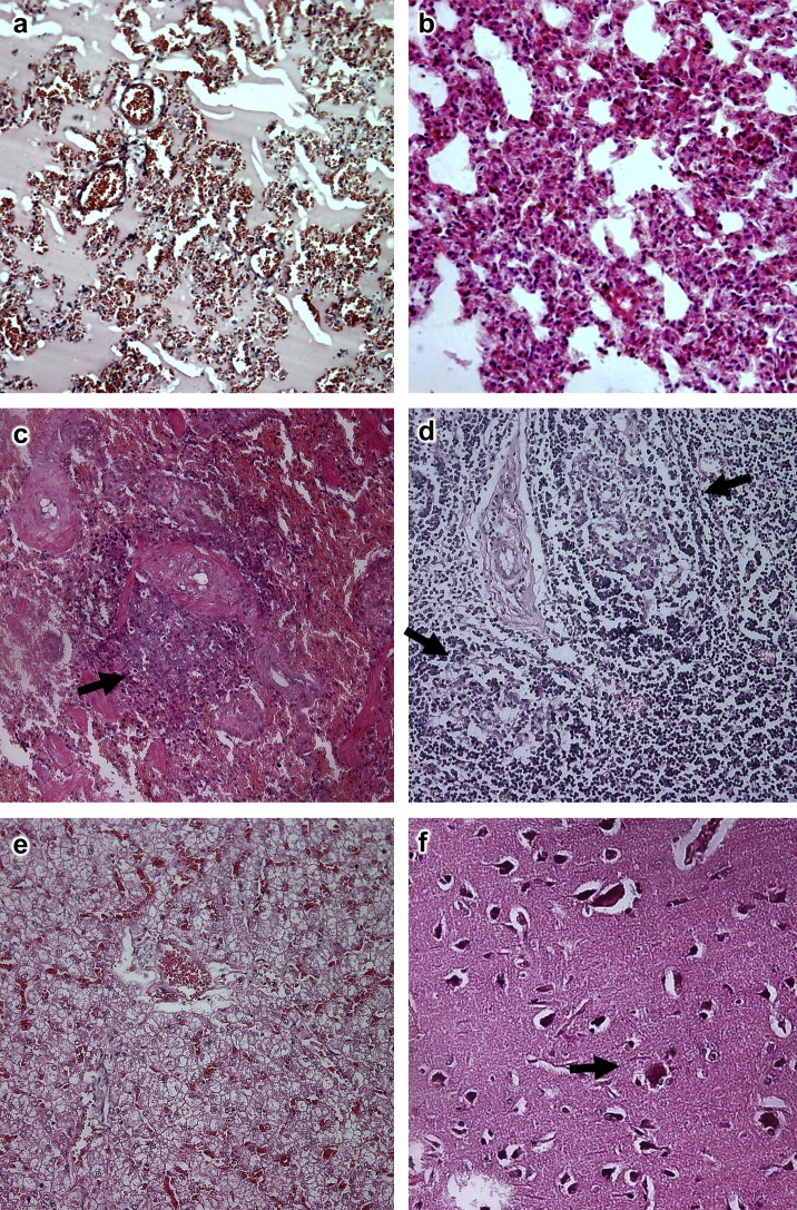
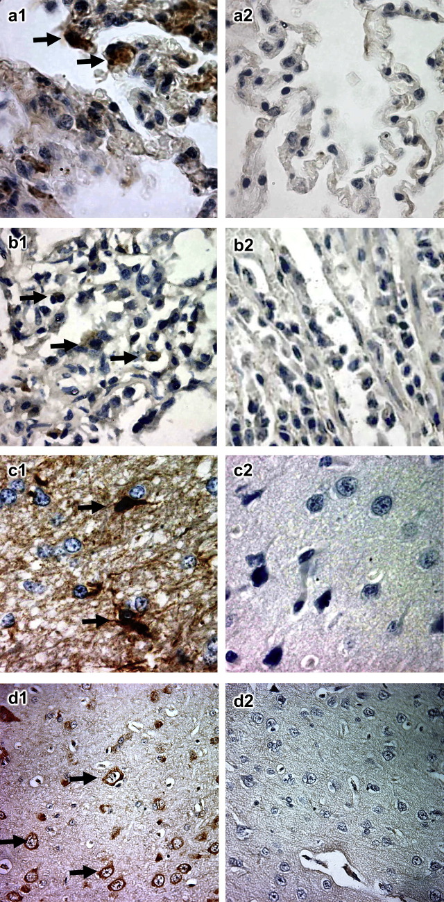
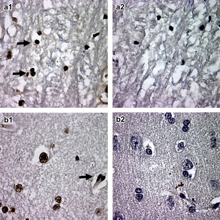
Masked palm civets are highly susceptible to infection with the severe acute respiratory syndrome coronavirus (SARS-CoV). Infected animals become less aggressive and develop pyrexia, lethargy and diarrhoea. The present study describes the spectrum of histopathological changes in the lung, spleen, lymph node, liver, small intestine, kidney and cerebrum of civets infected experimentally with SARS-CoV. In-situ hybridization (ISH) with probes specific for the RNA polymerase gene demonstrated viral RNA in the lung, small intestine and cerebrum only. In-situ labelling was employed in order to demonstrate cellular apoptosis in the cerebrum, but there was no evidence of apoptosis within the myocardium. These results indicate that SARS-CoV causes multi-organ pathology in civets, similar to that observed in human SARS patients. These parallels suggest that civets may be used as an animal model of this infection to gain insight into the pathogenesis of SARS and for evaluation of candidate vaccines and antiviral drugs.
Figures
Similar articles
-
Civets are equally susceptible to experimental infection by two different severe acute respiratory syndrome coronavirus isolates.J Virol. 2005 Feb;79(4):2620-5. doi: 10.1128/JVI.79.4.2620-2625.2005. J Virol. 2005. PMID: 15681462 Free PMC article.
-
A review of studies on animal reservoirs of the SARS coronavirus.Virus Res. 2008 Apr;133(1):74-87. doi: 10.1016/j.virusres.2007.03.012. Epub 2007 Apr 23. Virus Res. 2008. PMID: 17451830 Free PMC article. Review.
-
Organ distribution of severe acute respiratory syndrome (SARS) associated coronavirus (SARS-CoV) in SARS patients: implications for pathogenesis and virus transmission pathways.J Pathol. 2004 Jun;203(2):622-30. doi: 10.1002/path.1560. J Pathol. 2004. PMID: 15141376 Free PMC article.
-
Pneumonitis and multi-organ system disease in common marmosets (Callithrix jacchus) infected with the severe acute respiratory syndrome-associated coronavirus.Am J Pathol. 2005 Aug;167(2):455-63. doi: 10.1016/S0002-9440(10)62989-6. Am J Pathol. 2005. PMID: 16049331 Free PMC article.
-
Bats, civets and the emergence of SARS.Curr Top Microbiol Immunol. 2007;315:325-44. doi: 10.1007/978-3-540-70962-6_13. Curr Top Microbiol Immunol. 2007. PMID: 17848070 Free PMC article. Review.
Cited by
-
Coronavirus Infections of Animals: Future Risks to Humans.Biol Bull Russ Acad Sci. 2021;48(1):26-37. doi: 10.1134/S1062359021010052. Epub 2021 Mar 1. Biol Bull Russ Acad Sci. 2021. PMID: 33679117 Free PMC article.
-
SARS vaccines: where are we?Expert Rev Vaccines. 2009 Jul;8(7):887-98. doi: 10.1586/erv.09.43. Expert Rev Vaccines. 2009. PMID: 19538115 Free PMC article. Review.
-
Lessons from the host defences of bats, a unique viral reservoir.Nature. 2021 Jan;589(7842):363-370. doi: 10.1038/s41586-020-03128-0. Epub 2021 Jan 20. Nature. 2021. PMID: 33473223 Review.
-
Unraveling the Possible Routes of SARS-COV-2 Invasion into the Central Nervous System.Curr Treat Options Neurol. 2020;22(11):37. doi: 10.1007/s11940-020-00647-z. Epub 2020 Sep 25. Curr Treat Options Neurol. 2020. PMID: 32994698 Free PMC article. Review.
-
SARS-CoV nucleocapsid protein interacts with cellular pyruvate kinase protein and inhibits its activity.Arch Virol. 2012 Apr;157(4):635-45. doi: 10.1007/s00705-011-1221-7. Epub 2012 Jan 6. Arch Virol. 2012. PMID: 22222284 Free PMC article.
References
-
- Ding Y., He L., Zhang Q., Huang Z., Che X., Hou J., Wang H., Shen H., Qiu L., Li Z. Organ distribution of severe acute respiratory syndrome (SARS) associated coronavirus (SARS-CoV) in SARS patients: implications for pathogenesis and virus transmission pathways. Journal of Pathology. 2004;203:622–630. - PMC - PubMed
-
- Greenough T.C., Carville A., Coderre J., Somasundaran M., Sullivan J.L., Luzuriaga K., Mansfield K. Pneumonitis and multi-organ system disease in common marmosets (Callithrix jacchus) infected with the severe acute respiratory syndrome-associated coronavirus. American Journal of Pathology. 2005;167:455–463. - PMC - PubMed
Publication types
MeSH terms
Substances
LinkOut - more resources
Full Text Sources
Miscellaneous
